Corrections:
Correction: MYCN expression induces replication stress and sensitivity to PARP inhibition in neuroblastoma
Metrics: PDF 2184 views | ?
1Academic Unit of Molecular Oncology, Sheffield Institute for Nucleic Acids (SInFoNiA), Department of Oncology and Metabolism, University of Sheffield, Sheffield, UK
2Divisions of Clinical Studies and Cancer Therapeutics, The Institute of Cancer Research, Sutton, UK
3Divisions of Radiotherapy & Imaging, The Institute of Cancer Research, Sutton, UK
4The Children and Young People’s Unit, The Royal Marsden NHS Trust, Sutton, UK
5CRUK Gene Function Laboratory and Breast Cancer Now Research Centre, The Institute of Cancer Research, London, UK
6Sheffield Children’s Hospital, Western Bank, Sheffield, UK
#Present address: Medical Research Council Cancer Unit, Hutchison/Medical Research Council Research Centre, University of Cambridge, Cambridge, UK
*These authors contributed equally to this work
Published: January 12, 2023
Copyright: © 2023 King et al. This is an open access article distributed under the terms of the Creative Commons Attribution License (CC BY 4.0), which permits unrestricted use, distribution, and reproduction in any medium, provided the original author and source are credited.
This article has been corrected: In Supplementary Figure 2, under the IMR-32 column, the 3rd row, first panel image is an accidental duplicate of the 2nd row, 3rd panel image. The 6th row, 2nd panel image is also an accidental duplicate of the 7th row, 2nd panel image. In Supplementary Figure 11A, the 1st row, 2nd panel image was accidentally lifted from a student report as an example of IMR32 cells treated with DMSO. The corrected figures, obtained using the original data, are shown below. The authors declare that these corrections do not change the results or conclusions of this paper.
Original article: Oncotarget. 2020; 11:2141–2159. DOI: https://doi.org/10.18632/oncotarget.27329
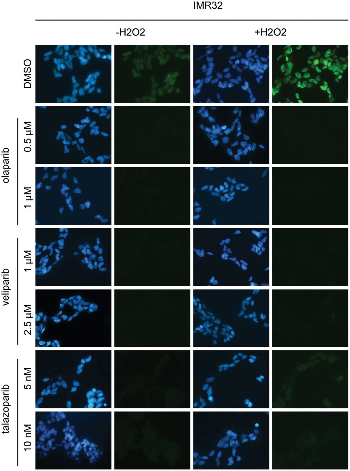
Supplementary Figure 2: PARP inhibitors olaparib, veliparib and talazoparib inhibit PARP in NB cells. Detection of PAR by immunofluorescence in IMR-32 and Shep-1 NB cell lines. Cells were pre-treated for 16 hours at the concentrations of PARP inhibitors as indicated. Treating cells with 150 μM H2O2 resulted in much stronger PAR activity in vehicle-treated cells and allowed PARP inhibition to be more clearly demonstrated. PAR (green), DAPI (blue).
 All site content, except where otherwise noted, is licensed under a Creative Commons Attribution 4.0 License.
PII: 28336