
Corrections:
Correction: PSC-derived Galectin-1 inducing epithelial-mesenchymal transition of pancreatic ductal adenocarcinoma cells by activating the NF-κB pathway
Metrics: PDF 1344 views | ?
1 Department of General Surgery, Institute of General Surgery, Northern Jiangsu Province Hospital, Clinical Medical College, Yangzhou University, Yangzhou, P.R. China
2 Department of General Surgery, Anhui No. 2 Provincial People’s Hospital, Hefei, Anhui Province, P.R. China
3 Department of General Surgery, The First Affiliated Hospital of Nanjing Medical University, Nanjing, P.R. China
4 Department of Clinical Medicine, Medical College of Yangzhou University, Yangzhou, P.R. China
* These authors have contributed equally to this work
Published: September 28, 2021
Copyright: © 2021 Tang et al. This is an open access article distributed under the terms of the Creative Commons Attribution License (CC BY 4.0), which permits unrestricted use, distribution, and reproduction in any medium, provided the original author and source are credited.
This article has been corrected: Due to accidental placement, some of the images in Figures 2 and 4 are incorrect. In Figure 2B, panels 2 and 3 have been replaced. In Figure 4C, row 3, all four panels have been replaced. The corrected Figures 2 and 4, produced using the original data, are shown below. The authors declare that these corrections do not change the results or conclusions of this paper.
Original article: Oncotarget. 2017; 8:86488–86502. DOI: https://doi.org/10.18632/oncotarget.21212
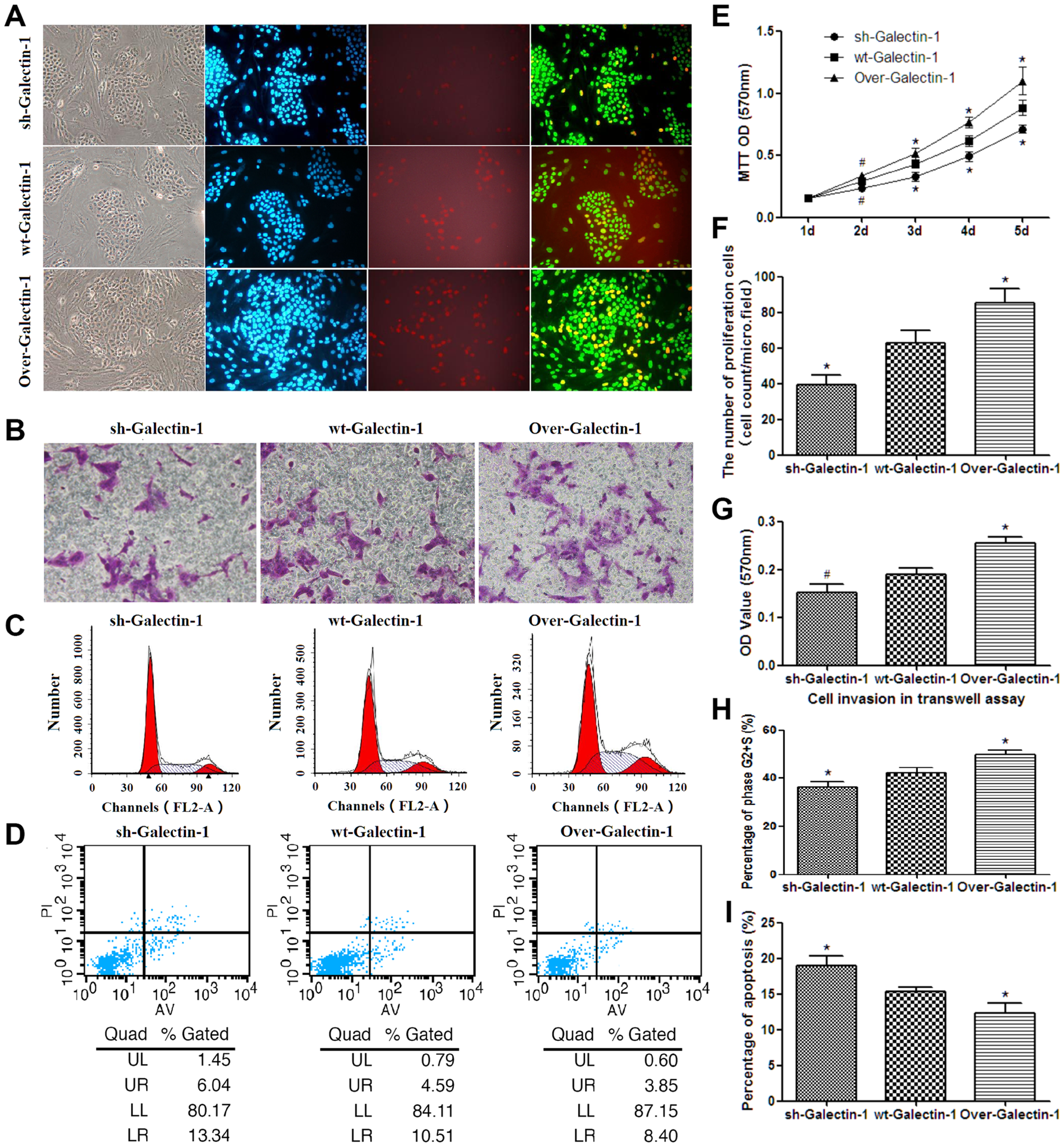
Figure 2: Effect of PSC-derived Galectin-1 on the proliferation and migration ability of PANC-1. (A) Proliferating capability of PANC-1 promoted by PSCs as evaluated by EdU incorporation (A). The number of proliferating cells (EdU cell count per micro field) is shown in (F) and cell growth over 5 days (measured using MTT assays) is shown in (E). (B) Invasion ability of PANC-1 promoted by PSCs as detected by the transwell invasion assay. The OD value of each group of invaded PANC-1 cells is shown in (G). (C) PSCs derived Galectin-1 promotes the proliferative activity (G2+S-phase fraction) of PANC-1 cells. Bar-graph representation of the G2+S-phase fraction cells in each group is shown in (H). (D, I) PSCs derived Galectin-1 has an anti-apoptotic effect on PANC-1 in vitro. All experiments were repeated three times. *p < 0.05, **p < 0.01, #p > 0.05 vs. wt-Galectin-1 PSCs.
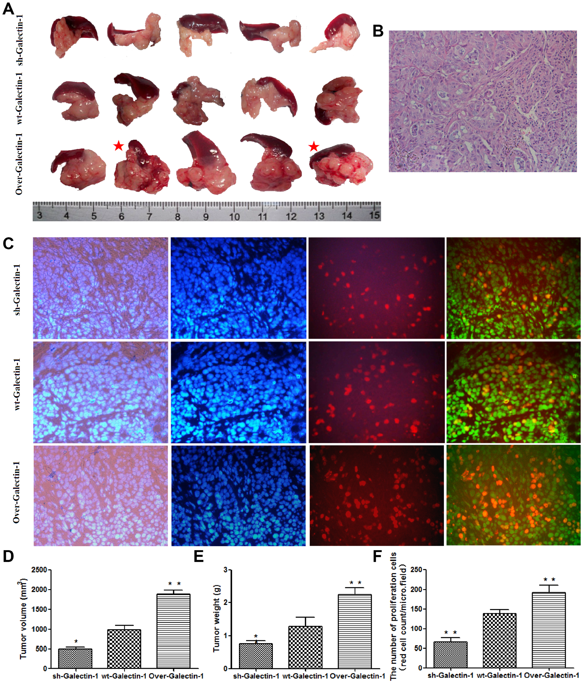
Figure 4: Effect of PSC-derived Galectin-1 on in vivo orthotopic xenograft establishment and growth. (A) PANC-1 mixed with PSCs were implanted orthotopically into the pancreas of nude mice (n = 5). The mice were sacrificed and the xenografts were removed on day 30 after cell implantation. The red star represent cases with liver metastasis. (B) H&E staining of samples of orthotopic xenografts in the pancreas of nude mice. (C) Proliferating capability of the orthotopic xenograft was evaluated using the EdU incorporation assay, and the number of EdU positive cells per micro field is shown in (F). Tumor volume (D) and weight (E) is expressed as the mean ± SE. *p < 0.05, **p < 0.01, #p > 0.05 vs. wt-Galectin-1 PSCs.
 All site content, except where otherwise noted, is licensed under a Creative Commons Attribution 4.0 License.
PII: 28087