Corrections:
Correction: Cbl-b inhibits P-gp transporter function by preventing its translocation into caveolae in multiple drug-resistant gastric and breast cancers
Metrics: PDF 1673 views | ?
1 Department of Medical Oncology, the First Hospital of China Medical University, Shenyang 110001, China
2 Department of Surgical Oncology and General Surgery, the First Hospital of China Medical University, Shenyang 110001, China
3 Department of Medical Respiratory, the First Hospital of China Medical University, Shenyang 110001, China
Published: August 03, 2021
Copyright: © 2021 Zhang et al. This is an open access article distributed under the terms of the Creative Commons Attribution License (CC BY 4.0), which permits unrestricted use, distribution, and reproduction in any medium, provided the original author and source are credited.
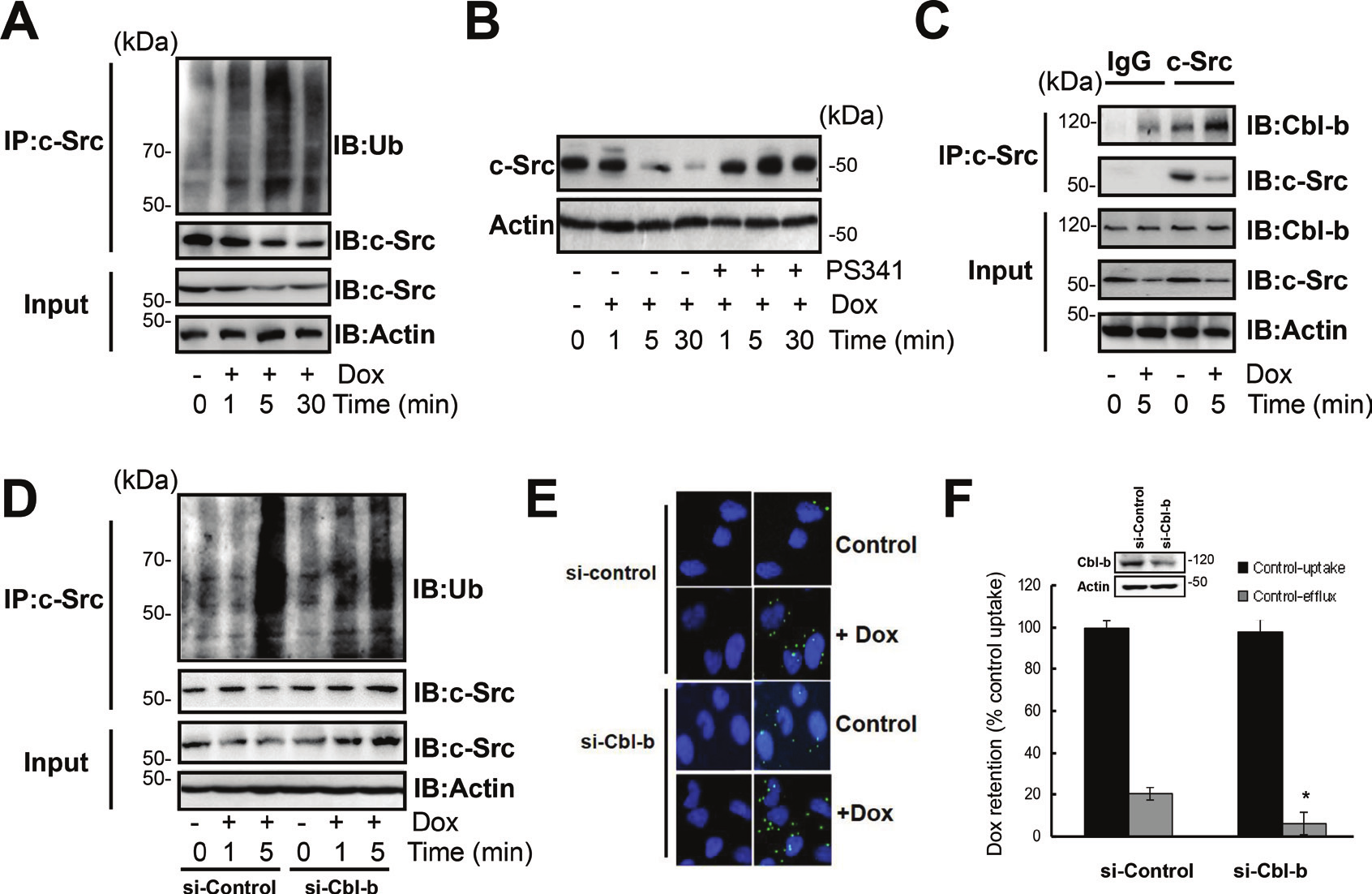
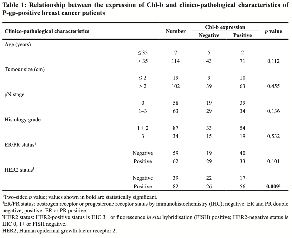
This article has been corrected: Due to errors during figure assembly, the fluorescence images in Figure 3E are accidental duplicates of those in Figures 1D and 2D. The corrected Figure 3, as well as an updated Figure 2 showing a correctly paired “control” and “+ DOX”, are shown below. In addition, the title of Table 1 should be “breast cancer”, not “gastric cancer.” All revisions presented were obtained with the original data. The authors declare that these corrections do not change the results or conclusions of this paper.
Original article: Oncotarget. 2015; 6:6737–6748. DOI: https://doi.org/10.18632/oncotarget.3253.

Figure 2: c-Src dependent Cav-1 phosphorylation promoted the translocation of P-gp into caveolae. (A) SGC7901/Adr cells were treated with or without 20 μg/ml Dox for 1, 5, 30, 60 min and the expression of P-gp, p-Src, Src, p-Cav-1, Cav-1 and Actin was detected by western blotting. (B) Cells were incubated with the Src inhibitor PP2 (10 μmol/l) for 2 h, treated with 20 μg/ml Dox for 5 min, and the expression of P-gp, p-Src, Src, p-Cav-1, Cav-1 and Actin was detected by western blotting. (C) SGC7901/Adr cells were pretreated with 10 μmol/l PP2 for 2 h followed by Dox treatment, and P-gp was immunoprecipitated and Cav-1 was analyzed western blotting. (D) In situ PLA in SGC7901/Adr cells pretreated with or without 10 μmol/l PP2 for 2 h, and then incubated with 20 μg/ml Dox for 5 min. Primary mouse and rabbit antibodies against P-gp and Cav-1 were combined with secondary PLA probes. (E) SGC7901/Adr cells were pretreated with 10 μmol/l PP2 for 2 h, followed by 20 μg/ml Dox and assessment of R-123 uptake and efflux.

Figure 3: Cbl-b inhibited the translocation of P-gp into caveolae by inducing the ubiquitination and degradation of c-Src. PFGE analysis of LCLs generated with recombinant B95-8, M81, or B95-8/npcEBNA1 virus. Samples are run as technical replicates for two independent donor generated LCLs. Cellular α-satellite DNA is shown as loading control above each lane. B. Quantitation of EBV episomes relative to α-satellite DNA for PFGE shown in panel A. C. Southern blot analysis of EBV terminal repeats after digestion with BamHI for B95-8, M81, or B95-8/npcEBNA1 generated LCLs. D. Western blot for EAD, ZTA, EBNA1, EBNA2, and Actin for B95-8, M81, or B95-8/npcEBNA1 generated LCLs.

 All site content, except where otherwise noted, is licensed under a Creative Commons Attribution 4.0 License.
PII: 27885
