Corrections:
Correction: miR146a-mediated targeting of FANCM during inflammation compromises genome integrity
Metrics: PDF 1662 views | ?
1 Laboratory of Genomic Instability and Cancer Therapeutics, Cancer Mutation Research Center, Chosun University School of Medicine, Gwangju, Republic of Korea
2 Laboratory of Genomic Instability and Cancer Therapeutics, Cancer Mutation Research Center, Chosun University School of Medicine, Gwangju, Republic of Korea
3 Department of Cellular and Molecular Medicine, Chosun University School of Medicine, Gwangju, Republic of Korea
4 Department of Oral Biology, Department of Applied Life Science, The Graduate School, Yonsei University College of Dentistry, Seoul, Republic of Korea
5 Department of Cell Biology, Microbiology, and Molecular Biology, College of Arts and Sciences, University of South Florida, Tampa, Florida, United States of America
* These authors contribute equally to this work
Published: May 26, 2020
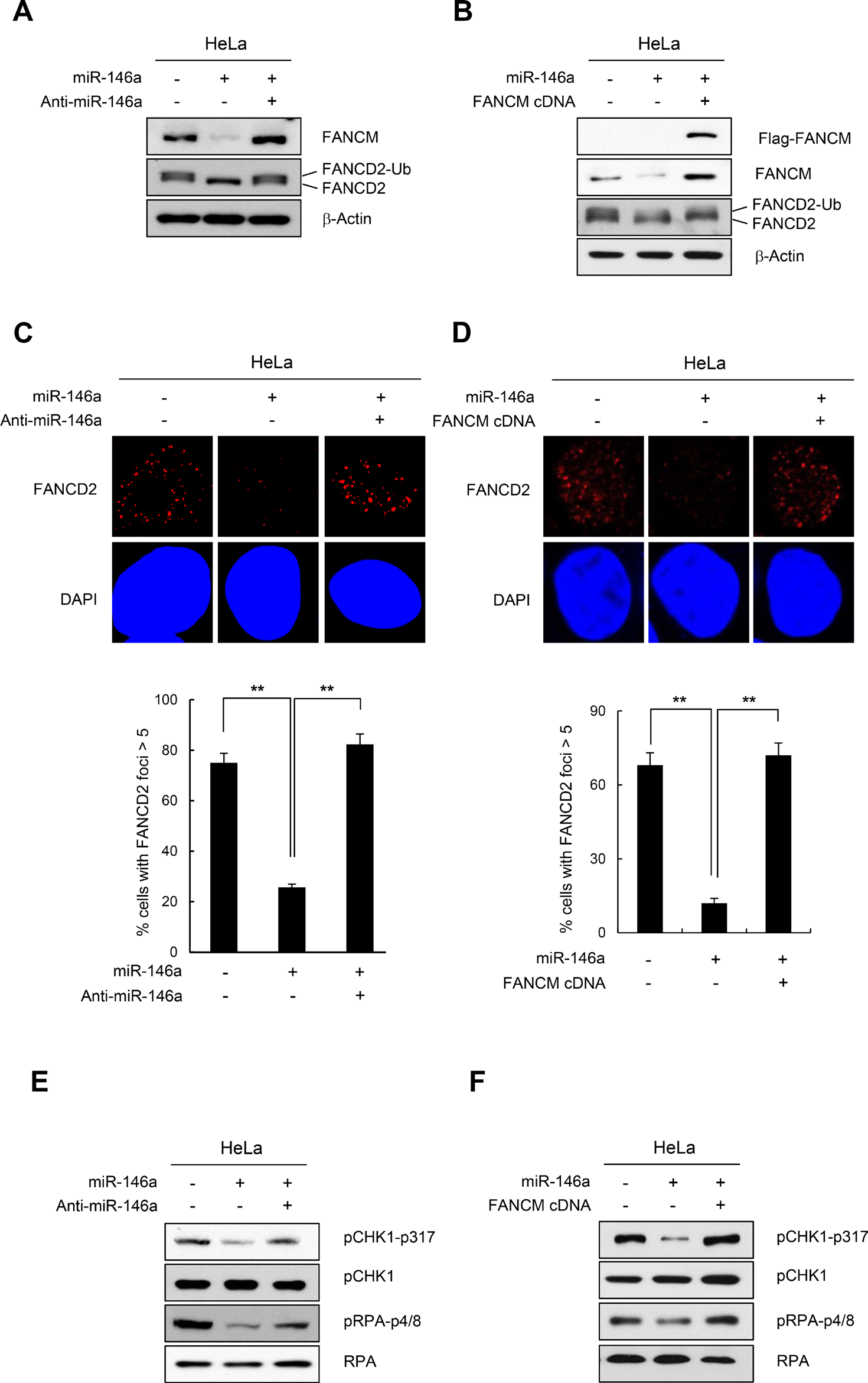
This article has been corrected: During the assembly of Figure 3E and 3F, the western blot of pCHK1-p317 in Figure 3F was inadvertently used for pRPA-p4/8 in Figure 3E. The corrected Figure 3 is shown below. The authors declare that these corrections do not change the results or conclusions of this paper.
Original article: Oncotarget. 2016; 7:45976–45994. DOI: https://doi.org/10.18632/oncotarget.10275.

Figure 3: Effect of miR146a on FANCD2 monoubiquitination and the FA pathway. (A and B) HeLa cells were transfected with miR146a in the absence or presence of anti-miR146a (A) or miR146a-insensitive FANCM cDNA (B). After a 48h transfection, the cells were treated with 5mM HU for 5 h. The protein levels of FANCM and FANCD2 were measured by western blotting. Monoubiquitinated FANCD2 is indicated as the upper band of doublet protein bands corresponding to FANCD2. (C and D) After HeLa cells underwent the same treatment as A and B, cells were analyzed for FANCD2 foci formation. DAPI was used for nuclear staining. Results are shown as the mean ± SD (n = 3); **P < 0.01. (E and F) Indicated cells were treated with 5mM HU for 5 hr. Cell lysates were analyzed by Western blotting with antibodies against pCHK1-S317, CHK1, pRPA-S4/8, and RPA.
 All site content, except where otherwise noted, is licensed under a Creative Commons Attribution 4.0 License.
PII: 27481
