Corrections:
Correction: Regulation of tumor suppressor EAF2 polyubiquitination by ELL1 and SIAH2 in prostate cancer cells
Metrics: PDF 1615 views | ?
1 Department of Urology, University of Pittsburgh School of Medicine, Pittsburgh, USA
2 Department of Pathology, University of Pittsburgh School of Medicine, Pittsburgh, USA
3 Department of Pharmacology and Chemical Biology, University of Pittsburgh School of Medicine, Pittsburgh, USA
4 University of Pittsburgh Cancer Institute, University of Pittsburgh School of Medicine, Pittsburgh, USA
5 Department of Geriatrics, Guangzhou General Hospital of Guangzhou Military Command, Guangzhou, China
6 Department of Urology, Shanghai General Hospital, Shanghai Jiao Tong University School of Medicine, Shanghai, China
7 Center for Translational Medicine, Guangxi Medical University, Nanning, Guangxi, China
8 Cancer Center, Traditional Chinese Medicine-Integrated Hospital, Southern Medical University, Guangzhou, China
9 Guangdong Provincial Key Laboratory of Geriatric Infection and Organ Function Support and Guangzhou Key Laboratory of Geriatric Infection and Organ Function Support, Guangzhou, China
Published: December 24, 2019
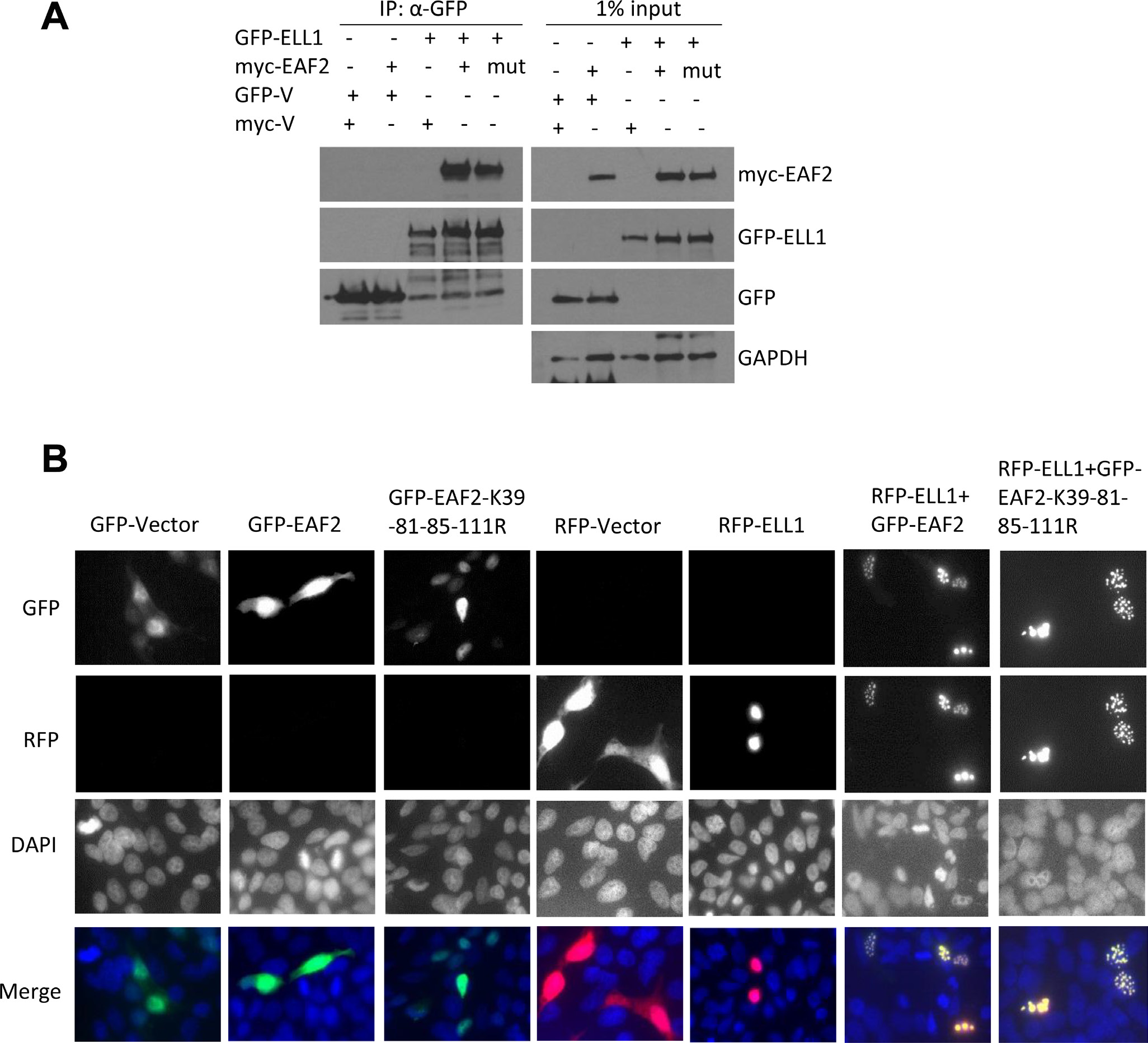
This article has been corrected: Due to errors during image assembly, the RFP-ELL1 merged image in Figure 4B is incorrect. The proper Figure 4 is shown below. The authors declare that these corrections do not change the results or conclusions of this paper.
Original article: Oncotarget. 2016; 7:29245–29254. DOI: https://doi.org/10.18632/oncotarget.8588.

Figure 4: Mutant EAF2K39-81-85-111R binding and co-localization with ELL1. (A) HEK 293 cells were transfected with myc-EAF2, myc-EAF2K39-81-85-111R, or empty myc expression vector together with GFP-ELL1 or empty GFP expression vector for 36 h. The cell lysates were prepared for co-immunoprecipitation using anti-GFP antibody. The precipitates and whole cell lysates (1% input) were analyzed by immunoblotting using anti-myc and anti-GFP antibodies. GAPDH in the whole cell lysates was probed as loading control. (B) C4-2 cells were transfected with GFP, GFP-EAF2, GFP-EAF2K39-81-85-111R, RFP, and RFP-ELL1 expression vector alone or in the indicated combinations for 48 h. Subcellular localization was imaged with confocal microscopy. Image enlargement: 100×. Data shown are representative of three independent experiments.
 All site content, except where otherwise noted, is licensed under a Creative Commons Attribution 4.0 License.
PII: 27354
