Corrections:
Correction: BNIP3 modulates the interface between B16-F10 melanoma cells and immune cells
Metrics: PDF 1837 views | ?
1 Laboratory for Cell Death Research and Therapy (CDRT), Department of Cellular and Molecular Medicine, KU Leuven, Leuven, Belgium
2 Laboratory of Hepatology, Department of Chronic Diseases, Metabolism and Ageing (CHROMETA), KU Leuven, Leuven, Belgium
3 Laboratory of Clinical and Experimental Endocrinology (CEE), Department of Chronic Diseases, Metabolism and Ageing (CHROMETA), KU Leuven, Leuven, Belgium
Published: December 11, 2018
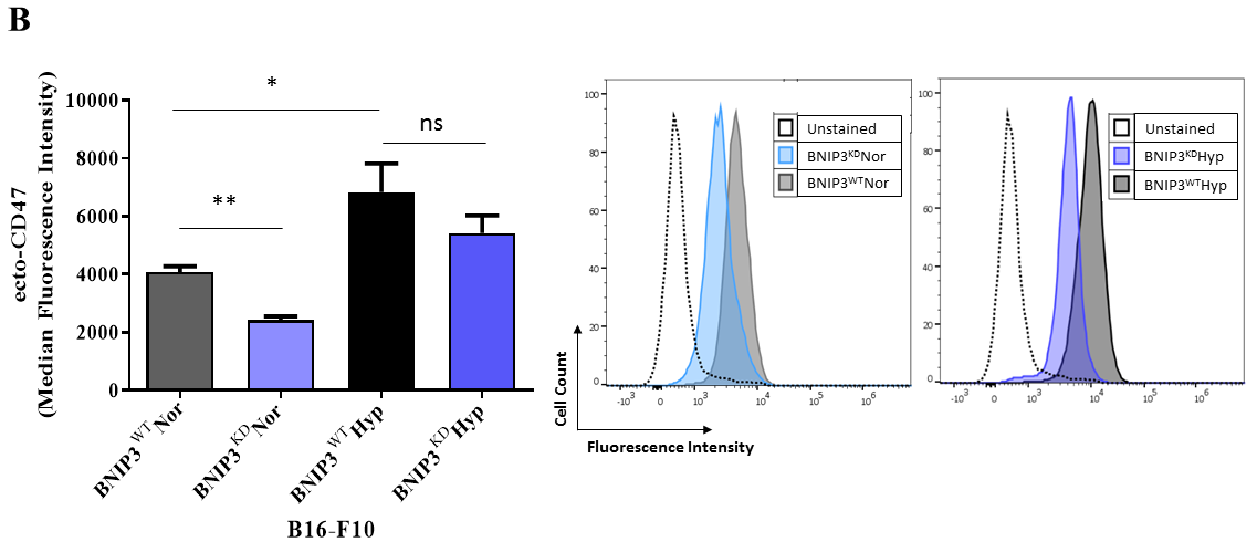
This article has been corrected: During the assembly of the Figure 1B, the flow cytometry histogram concerning ecto-CD47 expression of B16-F10 cell lines exposed to normoxic (Nor) conditions was presented incorrectly. Using the source data, a correct Figure 1B was generated and is shown below. The authors declare that these corrections do not change the results or conclusions of this paper.
Original article: Oncotarget. 2018; 9:17631-17644. DOI: https://doi.org/10.18632/oncotarget.24815.

Figure 1: BNIP3 and hypoxia modulate the phagocytosis of B16-F10 melanoma cells by macrophages. (B) Flow Cytometry- based quantification (left panel) and representative histograms (right panel) of the level of surface CD47 (ecto-CD47) in B16-F10 cells (BNIP3WT and BNIP3KD) after 24 h of culture in normoxia (Nor) or hypoxia (Hyp). Data expressed as mean ± SEM and analysed with Mann-Whitney’s t-test (**p = 0.0043, *p = 0.0411, ns = not significant [p = 0.3052]) as indicated by the bar, n = 3 independent experiments).
 All site content, except where otherwise noted, is licensed under a Creative Commons Attribution 4.0 License.
PII: 26474
