
INTRODUCTION
Digestive tract cancers (DTCs) are primary cancer burdens worldwide and include esophageal cancer (ESCA), gastric cancer (GC) and colorectal cancer (CRC), which are the ninth, sixth and third most common cancers in the world, respectively [1]. The incidence of DTCs has increased due to aging and growing populations and to increasing smoking and obesity rates [2, 3]. Although the diagnosis and treatment of DTCs have been improved for the past decades, the long-term survival rate for DTCs patients is still dismal and the cancer-related mortality rate is still high. Therefore, probing effective biomarkers for early diagnosis and clinical outcome prediction is imperative, which is helpful in better understanding the development of DTCs and further improving the survival outcome in patients with DTCs.
Protein p27, also known as CDKN1B (cyclin-dependent kinase inhibitor 1B), is a tumor suppressor protein of the CIP/KIP family and is encoded by p27 gene that located on chromosome 12p13. Protein p27 has been demonstrated to be a vital regulator of cell cycle progression via restraining cyclin E- and cyclin A-CDK2 complexes and preventing cells into S phase of the cell cycle [4]. Increasing researches have shown that p27 protein functions as a tumor suppressor to regulate both cell proliferation and tissue expansion in various malignancies. A growing number of studies have reported that p27 protein expression could be used to predict clinical outcomes in various malignant tumors, such as breast cancer [5], lung cancer [6] and ovarian cancer [7]. The clinical and prognostic values of p27 protein expression in patients with DTCs have also been investigated, with different studies reporting conflicting results; most studies have showed that a low p27 expression is related to a poor clinical outcome in DTCs patients [8, 9]. However, some contradictory results were also achieved [10, 11]. Thus. validation by independent studies working on different case series is urgently required before p27 protein can be served as a biomarker in clinic for diagnosis and clinical outcome prediction in patients DTCs. Therefore, we collected all available published literatures those referred to the clinical and prognostic significances of p27 protein in DTCs and performed immunohistochemistry (IHC) staining using clinical tissues from in-house DTCs patients to clarify the expression and clinical value of p27 protein in DTCs. Additionally, DTCs-related microarray and RNA-seq datasets were also obtained from the Gene Expression Omnibus (GEO), Oncomine, ArrayExpress and The Cancer Genome Atlas (TCGA) to further investigate the expression level and clinical significances of p27 gene in DTCs patients.
RESULTS
In the present study, IHC staining was conducted and relevant published literatures, microarray and RNA-seq datasets were collected to explore the expression patterns and clinical significances of p27 gene and protein in DTCs. The overall design of the current study is presented in Figure 1.
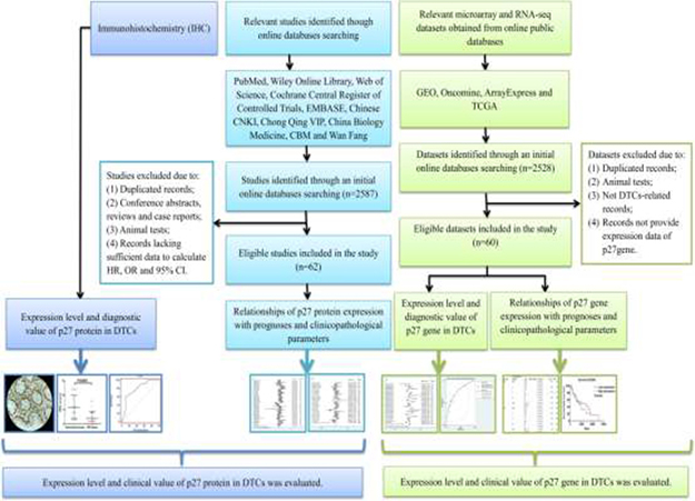
Figure 1: Flow chart of the overall design of the current study.
Expression level of p27 protein in DTCs based on IHC staining
Positive rate of p27 protein expression was 0% (0/5) in esophageal adenocarcinoma (ESAC) and 80% (4/5) in paired normal esophagus tissues, 0% (0/26) in esophageal squamous carcinoma (ESCC) and 26.9% (7/26) in paired normal esophagus tissues, 3% (1/32) in GC and 37.5% (12/32) in paired normal gastric tissues, 10% (3/30) in CRC and 50% (15/30) in paired normal colorectal cancer (Figure 2). The scatter diagram and paired T-test were utilized to compare the expression of p27 protein in DTCs tissues and adjacent non-cancerous tissues, and the results indicated that the expression level of p27 protein was lower in ESAC (P = 0.0078; Figure 3A), ESCC (P = 0.0002; Figure 3B), GC (P < 0.0001; Figure 3C) and CRC (P < 0.0001; Figure 3D) tissues than in paired non-tumorous tissues. The receiver operating characteristic (ROC) curve was generated to estimate the diagnostic capability of p27 protein in DTCs. The area under the curve (AUC) values of p27 protein in ESAC, ESCC, GC and CRC were 0.98 (95% CI: 0.66–1, P = 0.0001; sensitivity: 0.8; specificity: 1; Figure 3E), 0.79 (95% CI: 0.66–0.89, P = 0.0001; sensitivity: 0.69; specificity: 0.77; Figure 3F), 0.94 (CI: 0.86–0.99, P = 0.0001; sensitivity: 0.78; specificity: 1; Figure 3G) and 0.79 (95% CI: 0.67–0.88, P = 0.0001; sensitivity: 0.6; specificity: 0.9; Figure 3H), respectively.
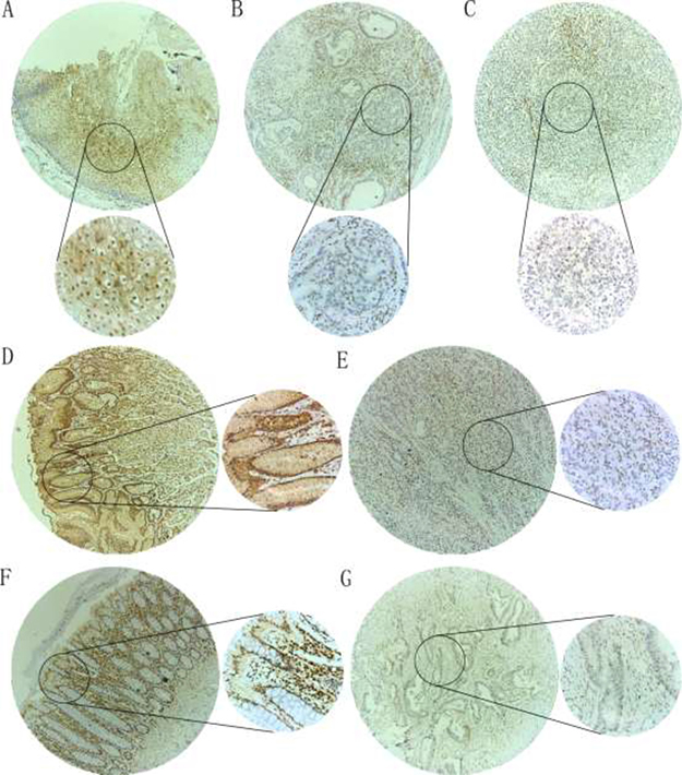
Figure 2: IHC staining of p27 protein in DTCs and adjacent non-tumorous tissues. Cytoplasmic and/or nuclear staining was observed for p27 protein. (A) A paracancerous esophagus tissue showed a high p27 protein expression (magnification: ×100 (upper) and ×400 (lower)); (B) A esophagus adenocarcinoma tissue showed a low p27 protein expression (magnification: ×100 (upper) and ×400 (lower)); (C) A esophagus squamous cell carcinoma tissue showed a low p27 protein expression (magnification: ×100 (upper) and ×400 (lower)); (D) A paracancerous gastric tissue showed a high p27 protein expression (magnification: ×100 (left) and ×400 (right)); (E) A gastric cancer tissue showed a low p27 protein expression (magnification: ×100 (left) and ×400 (right)); (F) A paracancerous colorectal tissue showed a high p27 protein expression (magnification: ×100 (left) and ×400 (right)); (G) A colorectal cancer tissue showed a low p27 protein expression (magnification: ×100 (left) and ×400 (right)).
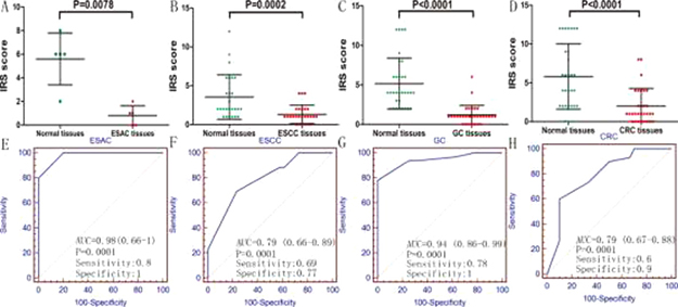
Figure 3: Expression level and diagnostic value of p27 protein in ESAC (A and E), ESCC (B and F), GC (C and G) and CRC (D and H).
Additionally, we also validated the expression level of p27 protein based on The Human Protein Atlas database (http://www.proteinatlas.org/), and the results are displayed in Supplementary Figure 1.
Clinical significances of p27 protein in DTCs based on published literatures
Result of literature search
As shown in Figure 1, a total of 62 publications (including 11 for ESCC, 26 GC for and 25 for CRC) with 9244 cases were involved in the present study. All of the literatures were published from 1997 to 2016. The expression of p27 protein was only assessed by regular IHC staining. The positive standards were inconsistent due to the use of different antibodies from different manufacturers. The follow-up times ranged from 29 to 240 months, the cut-off values ranged from 0.03 to 0.75, and the sample sizes ranged from 23 to 1062. Among the 62 studies, 54 studies [8, 10–62] followed survival, and 37 studies [8, 13, 15–20, 24, 28–30, 36, 39, 40, 44–47, 49–51, 54, 55, 57–61, 63–70] investigated the relationships of p27 protein expression with lymph node metastasis, distant metastasis and pathology grading. Among the 54 survival-related studies, 17 articles provided HRs and 95% CIs directly, while 33 articles only provided Kaplan–Meier survival curves. The basic information and quality scores of the 62 eligible studies are summarized in Supplementary Table 2.
Relationships of p27 protein expression with prognoses and clinicopathological parameters
A total of 48 reports with 8537 cases reported the correlation between p27 protein expression and overall survival (OS). The pooled hazard ratio (HR) from a random-effects model indicated that a low expression of p27 protein predicted a poor OS (HR: 1.58, 95% CI: 1.38–1.81, P < 0.0001; I2 = 69.2%, P < 0.0001; Figure 4; Table 1) in patients with DTCs. Then subgroup analyses according to cancer types, statistical methods and cut-off values were conducted. Results of subgroup analysis on cancer types showed that a down-regulated p27 protein indicated an unfavorable OS in patients with GC (HR: 1.47, 95% CI: 1.23–1.76, P < 0.0001; Supplementary Figure 2; Table 1) and CRC (HR: 1.81, 95% CI: 1.44–2.27, P < 0.0001; Supplementary Figure 2; Table 1), but not in patients with ESCC (HR: 1.41 95% CI: 0.92–2.14, P = 0.114; Supplementary Figure 2; Table 1). Subgroup analysis on statistical methods were also performed, and the HRs in multivariate analysis, univariate analysis and survival curve groups were 1.73 (95% CI: 1.27–2.34, P < 0.0001; Supplementary Figure 3; Table 1), 3.07 (95% CI: 1.83–5.14, P < 0.0001; Supplementary Figure 3; Table 1) and 1.46 (95% CI: 1.28–1.68, P < 0.0001; Supplementary Figure 3; Table 1), respectively. Results of subgroup analysis on cut-off values demonstrated that a reduced p27 protein expression was remarkably linked with a worse OS regardless of high (>0.25) or low (≤0.25) cut-off values, and the HRs were 1.63 (95% CI: 1.33 -2.0, P < 0.0001) and 1.46 (95% CI: 1.21–1.76, P < 0.0001), respectively (Supplementary Figure 4, Table 1). Additionally, a total of 5 studies with 564 patients investigated the relationship between p27 protein expression with disease-free survival (DFS) and 4 studies with 960 patients explored the association of p27 protein expression with cancer-specific survival (CSS). However, no statistically significant difference between p27 protein expression and DFS (HR: 1.20 95% CI: 0.62–2.34, P = 0.594; I2 = 86.4%, P < 0.0001; Figure 5A; Table 1) or CSS (HR: 1.37 95% CI: 0.61–3.06, P = 0.445; I2 = 82%, P = 0.001; Figure 5B; Table 1) was observed.
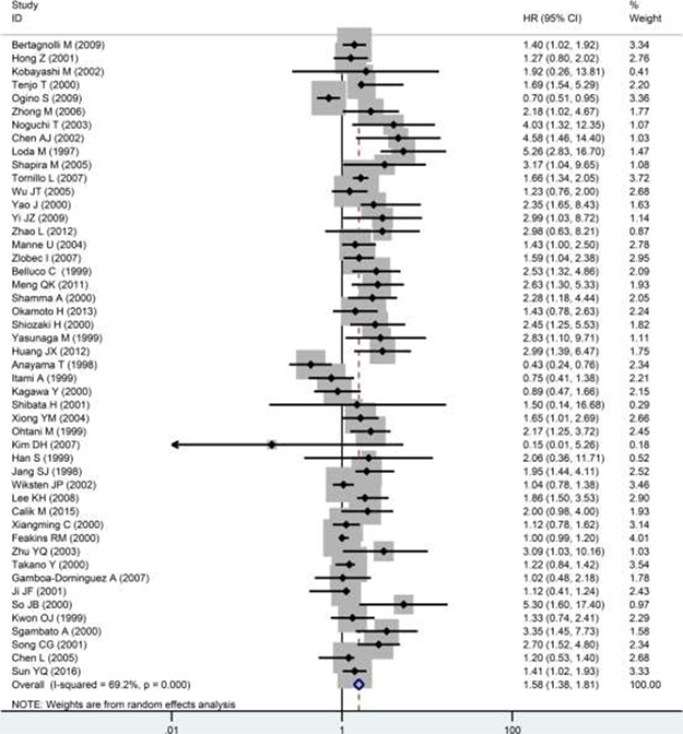
Figure 4: Forest plot of the pooled HR for OS. HR>1 indicates a poor OS for the group with a decreased p27 protein expression.
Table 1: The results of meta-analysis for OS, DFS and CSS
Survival outcome |
Group |
Number of studies |
Number of patients |
HR (95% CI) |
P value |
Heterogeneity test |
Publication bias |
||
|---|---|---|---|---|---|---|---|---|---|
I² (%) |
P value |
Begg’s P |
Egger’s P |
||||||
OS |
48 |
7853 |
1.58 (1.38–1.81) |
<0.0001 |
69.2 |
<0.0001 |
0.93 |
0.844 |
|
Cancer type |
|||||||||
ESCC |
10 |
773 |
1.41 (0.92–2.14) |
0.114 |
71.6 |
<0.0001 |
|||
GC |
19 |
2386 |
1.47 (1.23–1.76) |
<0.0001 |
66.1 |
<0.0001 |
|||
CRC |
19 |
4694 |
1.81 (1.44–2.27) |
<0.0001 |
64.9 |
<0.0001 |
|||
Statistical methods |
|||||||||
Multivariate analysis |
13 |
2374 |
1.73 (1.27–2.34) |
<0.0001 |
80.3 |
<0.0001 |
|||
Univariate analysis |
2 |
153 |
3.07 (1.83–5.14) |
<0.0001 |
0 |
0.318 |
|||
Survival curve |
33 |
5326 |
1.46 (1.28–1.68) |
<0.0001 |
48.4 |
0.001 |
|||
Cut-off value |
|||||||||
≤0.25 |
20 |
3551 |
1.46 (1.21–1.76) |
<0.0001 |
66.5 |
<0.0001 |
|||
>0.25 |
25 |
3849 |
1.63 (1.33–2.0) |
<0.0001 |
64.6 |
<0.0001 |
|||
DFS |
5 |
564 |
1.20 (0.62–2.34) |
0.594 |
86.4 |
<0.0001 |
None |
None |
|
CSS |
4 |
960 |
1.37 (0.61–3.06) |
0.445 |
82 |
0.001 |
None |
None |
|
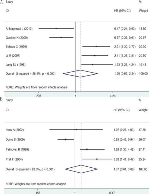
Figure 5: Forest plot of the pooled HR for DFS (A) and CSS (B).
Furthermore, the correlations of p27 protein expression with lymph node metastasis, distant metastasis and pathology grading were also assessed, and the results are concluded in Table 2. In total, 29 studies investigated the correlation between p27 protein expression and lymph node metastasis, 6 studies reported the relationship between p27 protein expression and distant metastasis and 25 studies evaluated the association of p27 protein expression with pathology grading. The overall odd ratios (ORs) indicated that a decreased expression of p27 protein was positively related to lymph node metastasis (OR: 2.15, 95% CI: 1.57–2.96, P < 0.0001; I2 = 69.4%, P < 0.0001; Figure 6A), distant metastasis (OR: 2.02, 95% CI: 1.12–3.63, P = 0.019; I2 = 50.4%, P = 0.073; Figure 6B) and pathology grading (OR: 2.14, 95% CI: 1.75–2.62, P < 0.0001; I2 = 17.2%, P = 0.221; Figure 7) in patients with DTCs. A subgroup analysis on cancer types was conducted. In ESCC group, a low expression of p27 protein was linked with pathology grading (OR: 2.16, 95% CI: 1.15–4.05, P = 0.017) but not with lymph node metastasis (OR: 1.97, 95% CI: 0.83–4.69, P = 0.124). In GC group, correlations of p27 protein expression with lymph node metastasis (OR: 2.05, 95% CI: 1.38–3.04, P < 0.0001) and pathology grading (OR: 2.08, 95% CI: 1.62–2.69, P < 0.0001) were observed; while no significant associations between p27 protein expression and distant metastasis was found (OR: 1.19, 95% CI: 0.74–1.91, P = 0.463). In CRC group, a down-regulated p27 protein was positively correlated with lymph node metastasis (OR: 2.79, 95% CI: 1.33–5.88, P = 0.007; Figure 6A), distant metastasis (OR: 3.92, 95% CI: 2.05–7.49, P < 0.0001) and pathology grading (OR: 2.28, 95% CI: 1.55–3.36, P < 0.0001).
Table 2: The relationships of p27 protein expression with lymph node metastasis, distant metastasis and pathology grading
Clinicopathological features |
Group |
Number of studies |
Number of patients |
OR (95% CI) |
P value |
Heterogeneity test |
Publication bias |
||
|---|---|---|---|---|---|---|---|---|---|
I² (%) |
P value |
Begg’s P |
Egger’s P |
||||||
Lymph node metastasis |
Overall result |
29 |
4208 |
2.15 (1.57–2.95) |
<0.0001 |
69.4 |
<0.0001 |
0.586 |
0.611 |
ESCC |
5 |
462 |
1.97 (0.83–4.69) |
0.124 |
65.4 |
0.021 |
|||
GC |
18 |
3100 |
2.05 (1.38–3.04) |
<0.0001 |
72.6 |
<0.0001 |
|||
CRC |
6 |
646 |
2.79 (1.33–5.88) |
0.007 |
64.8 |
0.014 |
|||
Distant metastasis |
Overall result |
6 |
921 |
2.02 (1.12–3.63) |
0.019 |
50.4 |
0.073 |
None |
None |
GC |
3 |
694 |
1.19 (0.74–1.91) |
0.463 |
0 |
0.967 |
|||
CRC |
3 |
247 |
3.92 (2.05–7.49) |
<0.0001 |
0 |
0.467 |
|||
Pathology grading |
Overall result |
25 |
2344 |
2.14 (1.75–2.62) |
<0.0001 |
17.2 |
0.221 |
0.907 |
0.911 |
ESCC |
4 |
270 |
2.16 (1.15–4.05) |
0.017 |
0 |
0.489 |
|||
GC |
11 |
1139 |
2.08 (1.62–2.69) |
<0.0001 |
32.3 |
0.141 |
|||
CRC |
10 |
935 |
2.28 (1.55–3.36) |
<0.0001 |
21.8 |
0.242 |
|||
Abbreviations: SD: standard deviation
Table 3: The expression level and diagnostic performance of p27 gene in DTCs
Gorup |
Number of datasets |
SMD (95% CI) |
P value |
Heterogeneity |
SROC curve |
|||
|---|---|---|---|---|---|---|---|---|
I² (%) |
P value |
AUC (95% CI) |
Sensitivity |
Specificity |
||||
Overall result |
60 |
0.10 (-0.08~0.27) |
0.274 |
83.7 |
<0.0001 |
0.79 (0.76–0.83) |
0.60 |
0.84 |
ESAC |
6 |
-0.56 (-1.55~0.44) |
0.273 |
89.9 |
<0.0001 |
0.82 (0.78–0.85) |
0.76 |
0.86 |
ESCC |
9 |
-0.45 (-0.74~-0.16) |
0.002 |
50.6 |
0.04 |
0.85 (0.82–0.88) |
0.69 |
0.84 |
GC |
20 |
-0.06 (-0.29~0.17) |
0.614 |
75.9 |
<0.0001 |
0.74 (0.70–0.78) |
0.53 |
0.82 |
CRC |
25 |
0.59 (0.34~0.83) |
<0.0001 |
79.2 |
<0.0001 |
0.81 (0.78–0.84) |
0.61 |
0.84 |
Abbreviations: SROC curve: summary receiver operating characteristic curve; AUC: area under the curve.
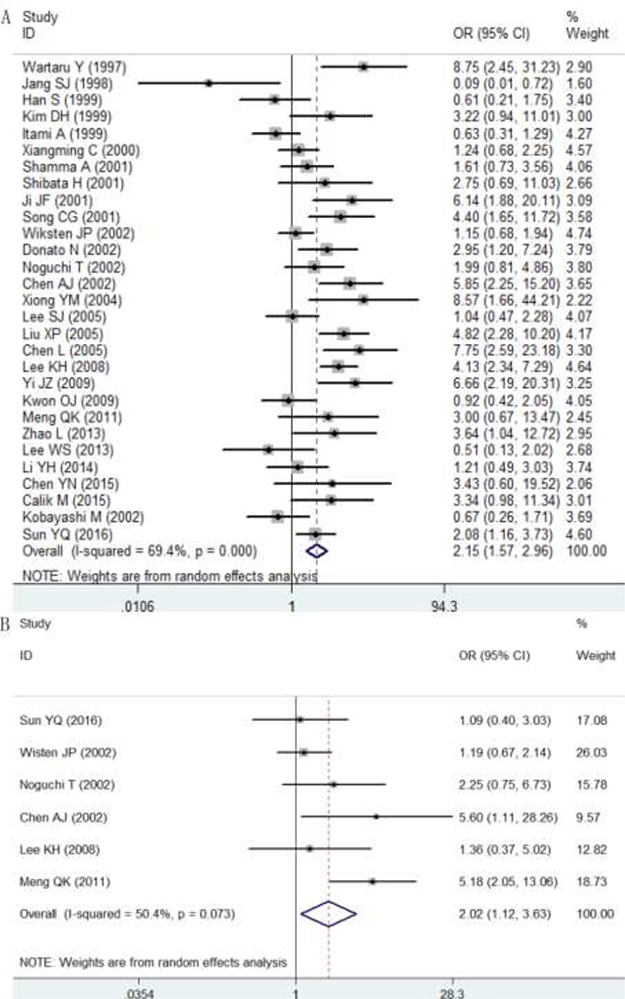
Figure 6: Relationships of p27 protein expression with lymph node metastasis (A) and distant metastasis (B). OR >1 indicates that a decreased p27 expression was positively correlated with lymph node metastasis and distant metastasis.
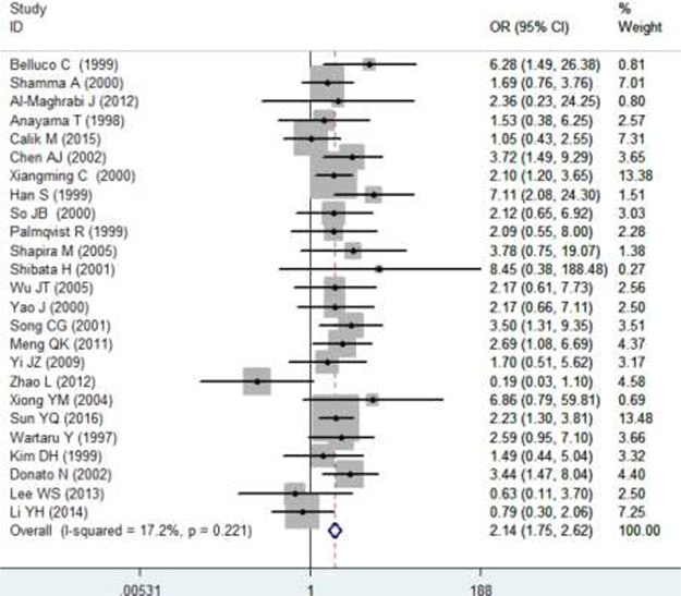
Figure 7: Relationships between p27 protein expression and pathology grading. OR>1 indicates that a decreased p27 expression was positively correlated with pathology grading.
Publication bias were also detected using Begg’s and Eeggr’s tests, and the results showed no publication bias in all situations (Table 1 and Table 2; Supplementary Figure 5).
Expression level and clinical significances of p27 gene in DTCs based on microarray and RNA-seq datasets
Expression level and diagnostic performances of p27 gene in DTCs
A total of 60 mRNA microarray and RNA-seq datasets including 6 for ESAC, 9 for ESCC, 20 for GC and 25 for CRC were mined in the present study. The essential information of the 60 datasets is showed in Supplementary Table 2.
To evaluate the expression level of p27 gene in DTCs, we integrated the 60 datasets. As shown in Table 3, the overall standard mean deviation (SMD) from a random-effects model was 0.1 (95% CI = -0.08~0.27, P = 0.274; I² = 83.7%, P < 0.0001; Figure 8), indicating that the expression level of p27 gene in DTCs was similar to that in normal controls. Then a subgroup analysis on cancer types was performed. The results showed that p27 gene was down-regulated in ESCC (SMD = -0.45, 95% CI = -0.74~-0.16, P = 0.002; I² = 50.6%, P = 0.04; Figure 9A) and up-regulated in CRC (SMD = 59, 95% CI = 0.34~0.83, P < 0.0001; I² = 79.2%, P < 0.0001; Figure 9B). While the pooled SMD for ESAC (SMD = -0.56, 95% CI = -1.55~0.44 P = 0.273; I² = 89.9%, P < 0.0001; Figure 10A) and GC (SMD = -0.06, 95% CI = -0.29~0.17, P = 0.614; I² = 75.9%, P < 0.0001; Figure 10B) suggested no statistically significant difference between cancerous and adjacent non-cancerous tissues. Additionally, the expression pattern of p27 gene in DTCs based on TCGA datasets were also displayed in the form of scatter plots and ROC curves (Supplementary Figure 6).
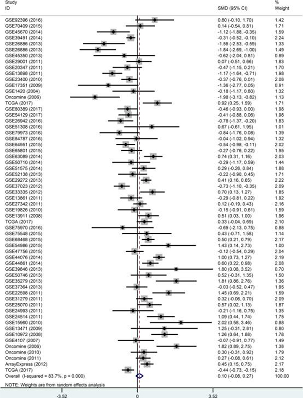
Figure 8: Forest plot of the 60 datasets evaluating p27 gene expression in DTCs (random-effects model). SMD >0 indicates that p27 gene was up-regulated in DTCs.
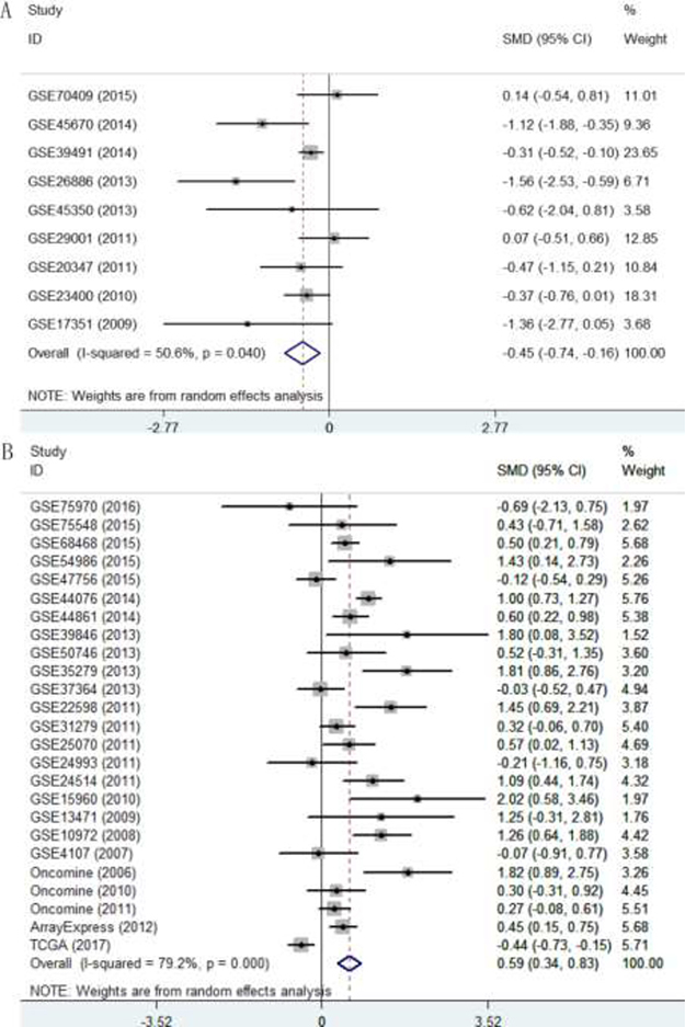
Figure 9: Forest plot evaluating p27 gene expression in ESCC and CRC. (A) Expression level of p27 gene in ESCC. (B) Expression level of p27 gene in CRC.
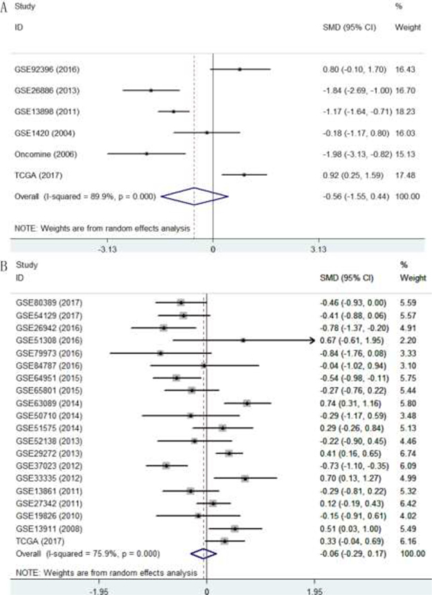
Figure 10: Forest plot evaluating p27 gene expression in ESAC and GC. (A) Expression level of p27 gene in ESAC; (B) Expression level of p27 gene in GC.
Begg’s and Egger’s tests were conducted to estimate the publication bias among the 60 datasets, and the results demonstrated no publication bias (Begg’s P = 0.934, Egger’s P = 0.923; Supplementary Figure 7).
Furthermore, the summary receiver operating characteristic (SROC) curve was also generated to assess the ability of p27 in discriminating DTCs from normal controls. As shown in Table 3, the overall AUC of p27 gene in DTCs was 0.79 (95% CI: 0.76–0.83; Figure 11A), with sensitivity and specificity of 0.60 and 0.84, respectively. In addition, the AUC values of p27 gene in ESAC, ESCC, GC and CRC were 0.82 (CI: 0.78–0.85; sensitivity: 0.76; specificity: 0.86; Figure 11B), 0.85 (CI: 0.82–0.88; sensitivity: 0.69; specificity: 0.84; Figure 11C), 0.74 (CI: 0.70–0.78; sensitivity: 0.53; specificity: 0.82; Figure 11D) and 0.81 (CI: 0.78–0.84; sensitivity: 0.61; specificity: 0.84; Figure 11E), respectively.
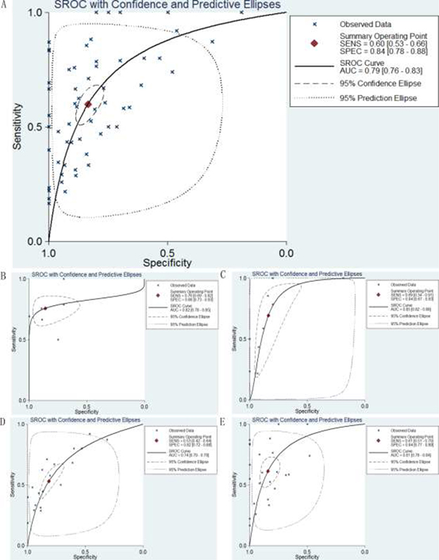
Figure 11: SROC curves for the identification of DTCs patients from normal controls using p27 gene expression. (A) Diagnostic performance of p27 gene in DTCs; (B) Diagnostic performance of p27 gene in ESAC; (C) Diagnostic performance of p27 gene in ESCC; (D) Diagnostic performance of p27 gene in GC; (E) Diagnostic performance of p27 gene in CRC.
Clinical significances of p27 gene in DTCs from TCGA data
Expression data of p27 gene and corresponding clinicopathological information were downloaded from TCGA to explore the clinical value of p27 gene in DTCs. The correlations between p27 gene expression and several key clinicopathological parameters, including pathology grading, T stage, N stage, M stage and TNM stage, were assessed using Independent-samples t-test. The prognostic values of p27 gene in DTCs were assessed using Log-Rank test and Kaplan–Meier survival curve. However, according to our results, no significant associations of p27 gene expression with those key clinicopathological parameters and prognoses were observed (Supplementary Table 1 and Figure 8).
DISCUSSION
The basic features of cancer are the disordered and uncontrolled cell cycle and the unlimited cell proliferation. As a member of cyclin-dependent kinase inhibitor family, p27 protein acts as a negative controller of cellular proliferation through inducing G1 arrest of the cell cycle, and thus inhibits the occurrence and development of malignant tumors [71–74]. A large number of researches have showed that the expression level of protein p27 in cancerous tissues is lower than that in normal controls, indicating that p27 protein may function as a tumor suppressor in malignancies. In our present study, we also detected the expression pattern of p27 protein in DTCs and adjacent non-cancerous tissues using IHC. Our results showed that p27 protein was down-regulated in ESAC, ESCC, GC and CRC, which are consistent with preceding researches [14, 25, 49, 75]. Then we further investigated the diagnostic ability of p27 protein in DTCs, and the results suggested that p27 protein could differentiate patients with DTCs from normal subjects to a degree. However, the results should be interpreted with caution since the number of the included in-house specimens is small. Therefore, further investigations with larger sample scales are necessary to verify our conclusion.
A study conducted by See et al [76] show that an increase in the number of chromatid breaks in protein p27-absence cells leads to a decrease in chromosome stability and ultimately a poor prognosis in cancer patients. Previous studies have also demonstrated that cancer patients with down-regulation of p27 protein are at high risk for poor clinical outcome [5, 7, 77, 78]. However, the clinical and prognostic value of protein p27 in DTCs has not been systematically investigated as of yet. Therefore, we collected all available relevant published literatures to synthetically analyze the relationships of p27 protein expression with prognoses and clinical parameters. A total of 54 publications were included to estimate the prognostic role of p27 protein in DTCs. The overall results indicated that the loss of p27 protein expression was an independent prognostic biomarker for OS but not for DFS and CSS in patients with DTCs. On the one hand, reports those investigated OS usually contained patient with longer follow-up time and included more cases to obtain statistical results on patient prognosis than those investigated DFS and CSS, and thus yielded stronger data. On the other hand, the number of included studies reporting DFS and CSS were only five and four, respectively. Thus, further studies with more samples are necessary to determine the correlations of p27 protein expression with DFS and CSS. Additionally, stratified analysis was also undertaken, and the results suggested that a decreased p27 protein expression in DTCs is predictive for OS independent of statistical method and cut-off value. However, subgroup analysis on cancer types showed that a down-regulated p27 protein predicted an unfavorable OS in GC and CRC but not in ESCC. A possible explanation is that the histologic type of ESCC is different from that of GC and CRC: CRC and GC are predominantly adenocarcinoma, while ESCC originates from squamous cells. The contributing manner of cell-cycle regulator p27 protein in ESCC is probably different from that in adenocarcinoma [17, 31, 79].
As well known, cancer patients with lymph node or distant metastasis often encounter poor survival outcome. Meanwhile, high grade of carcinoma pathology usually means poor tumor differentiation and prognosis. Therefore, digging an effective biomarker to predict cancer progression and identify high-risk patients, thereby optimizing individual treatment management and improving the prognosis of DTCs patients is imperative. According to our results, a reduced p27 protein expression in patients with DTCs is closely correlated with positive lymph node and distant metastases as well as poor tumor differentiation. Detection of p27 protein in patients with DTCs may be helpful in distinguishing high-risk patients and guiding the determination of individualized treatment protocols. Further and stricter clinical researches are needed to verify the conclusion.
Protein p27 is encoded by gene p27, while the cellular abundance of p27 protein is mainly regulated at the post-transcriptional level and the regulation is affected by a lot of factors. Accumulated evidences have demonstrated that phosphorylation induced by PKB/Akt [80, 81] or CyclinD1 [82] dominates the expression and stability of p27 protein. A study conducted by Loda et al [31] demonstrate that tumors with decreased p27 protein expression show increased proteolytic activity specific for protein p27, indicating that the down-regulated p27 protein may be due to the enhancive proteasome-induced degradation rather than the altered expression of p27 gene. To clarify the correlations between p27 protein and p27 gene in DTCs, we collected 61 microarray and RNA-seq datasets those contained the expression data of p27 gene. The overall result showed that the expression level of p27 gene in DTCs is similar to that in normal controls. Then a subgroup analysis by cancer types was also carried out, and the results indicated that p27 gene was down-regulated in ESCC but up-regulated in CRC. While in ESAC and GC, no altered expression of p27 gene was observed. Additionally, we also investigated the clinical and prognostic value of p27 gene in DTCs, and the results presented that no relationships of p27 gene expression with clinicopathological parameters and prognoses were found. Our findings suggested that there was no correlation between p27 protein and p27 gene expression in DTCs, which was in accordance with preceding reports conducted by Hunter et al [83] and Li et al [84].
Although our results revealed that p27 protein was down-regulated in DTCs and the reduced expression of p27 protein was closely related to poor clinical outcome, the conclusions should be interpreted cautiously since there were several limitations in our study. First, the combined HR used in our study induced a key bias due to the use of different statistical methods in the included studies, including multivariate analysis, univariate analysis and survival curves. The HRs acquired from multivariate analyses were more accurate than those from univariate analyses because intermixed factors were included in the multivariate analyses. Additionally, in several reports, the HRs were not provided directly and had to be calculated from the Kaplan–Meier survival curves, which may result in inevitable errors and decrease the accuracy of the results. Second, extensive significant heterogeneities were observed in our study. The noticeable heterogeneities may result from the differences in the following characteristics of included records: sample sizes, detection methods and platforms, primary antibodies and dilutions, cut-off values, preoperative treatments, cancer types, case selection criteria, population differences in regions, gender ratios, tumor stages, and status of lymph node and distant metastases. Third, the length of follow-up times in the included trials ranged from 29 months to 240 months, and the number of participants varied from 23 to 1062. We did not exclude studies with limited follow-up times or small patient numbers. However, this may introduced biases because the limited durations and study sizes are likely to produce unreliable data. Fourth, our study included fully published studies in English or Chinese and excluded studies written with other languages, which may introduced potential language bias. Additionally, researches with positive results are more likely to be published. Therefore, we should not ignore the potential bias in the present study even though the results from Begg’s and Egger’s tests showed no publication bias.
To sum up, our results suggested p27 protein was down-regulated in DTCs and a decreased expression of p27 protein predicted an unfavorable clinical outcome. Detecting p27 protein in patients with DTCs may be helpful in distinguishing high-risk patients and guiding the determination of individualized treatment protocols. More interesting, we also discovered that the expression level and clinical significances of p27 protein were inconsistent with those of p27 gene, indicating that the down-regulation of p27 protein did not result from the altered expression of p27 gene. Further well-designed studies are warranted to validate our conclusions.
MATERIALS AND METHODS
Immunohistochemistry
The expression level of p27 protein was detected by IHC method in 26 ESCC, 5 ESAC, 32 GC, 30 CRC and their adjacent normal tissues. All samples were obtained from patients who underwent surgery without neoadjuvant treatment at Guangxi Medical University, People’s Republic of China from May 2015 to April 2017. The formalin-fixed paraffin-embedded tissues were cut into 4-μm thick sections and deparaffinized. Antigen retrieval was performed in EDTA buffer with pressure cooking at 100°C for 4 minutes. Endogenous peroxidase was inactive in 3% hydrogen peroxide for 15 minutes. Then the sections were incubated with rabbit monoclonal anti-p27 KIP antibody (1:100 dilution, Abcam, USA) for 1 h at 37°C. The rest procedure was conducted according to the manufacturer’s instruction. Two pathologists (Gang Chen and Yi-wu Dang) read each immunostained tissue under microscopy and scored it independently according to the following two criteria: (1) the staining intensity was determined as 0 (no staining), 1 (weak staining), 2 (moderate staining) and 3 (strong staining); (2) the staining percentage of tumor cells was scored as 0 (<5%), 1 (5%–25%), 2 (26%–50%), 3 (51%–75%), 4 (76%–100%). The positive percentage of cytoplasm and/or nuclei of cells were calculated in more than 1000 cells of five successive and representative high power fields (×400 magnification microscope). The immunoreactive score (IRS) was applied to determine the final staining score by multiplication of the intensity score and the distribution score. Patients were divided into p27 positive group (IRS ≥ 6) and p27 negative group (IRS < 6). Then the scatter diagram was generated using GraphPad Prism 5 (La Jolla, CA, USA) and the SROC curve analysis was performed using Stata12.0 (Stata Corporation, College Station, TX, USA) based on the IRS scores to evaluate the expression pattern and diagnostic capability of p27 protein in DTCs.
Data mining
Data acquisition from published literature
A comprehensive and systematic literature search was conducted in 10 online databases, including Wiley Online Library, Web of Science, Cochrane Central Register of Controlled Trials, EMBASE, PubMed, Chinese CNKI, Chong Qing VIP, China Biology Medicine disc, CBM and Wan Fang (with an upper date limit of August 15, 2017), using the following strategy: (malignan* OR cancer OR tumor OR tumour OR neoplas* OR carcinoma OR adenocarcinoma) AND (digestive OR gastrointestinal OR gastric OR stomach OR esophageal OR esophagus OR gut OR intestinal OR colorectal OR colonic OR rectal OR colon OR rectum) AND (p27KIP1 OR KIP1 OR MEN4 OR MEN1B p27 OR CDKN4 OR “cyclin-dependent kinase inhibitor 1B” OR “cyclin dependent kinase inhibitor 1B” OR CDKN1B).
Relevant publcations were enrolled based on the following inclusion criteria: (1) patients were diagnosed with esophageal, gastric or colorectal cancer pathologically; (2) studies elaborated the relationships of p27 expression with prognoses or clinicopathological parameters; (3) studies were written in English or Chinese as full papers; (4) published data must be sufficient to determine the HR, OR and 95% CI; and (5) the most complete and recent article was selected when multiple articles based on the same patient set were found.
Papers were not considered based on the following exclusion criteria: (1) conference abstracts, case reports and reviews; (2) animal tests; and (3) records lacking sufficient data to calculate HR, OR and 95% CI.
Main characteristics of all of the included studies were extracted by two investigators (Dan-dan Xiong and Ai-hua Lan) independently to ensure homogeneity in information gathering and entry. The following information were gathered: first author, publication year, cancer type, cut-off value, country, sample size, follow-up period, statistical method, HR with corresponding 95% CI and study quality score. Each discrepancy was determined through discussion with a third reviewer (Gang Chen). The Newcastle-Ottawa quality assessment scale was utilized to estimate the quality of each study [85]. The HRs and 95% CIs were collected directly if they were explicitly provided in the original articles; otherwise, they were extracted from the Kaplan–Meier survival curves using Engauge Digitizer Version 4.1. Multivariate analysis can better reveal the influence of multiple factors on the survival response. Therefore, the multivariate HRs and 95% CIs were selected if multivariate and univariate analyses were available in the same study. Then we calculated the pooled HR to estimate the prognostic value of p27 in patients with DTCs. Furthermore, stratified analyses according to cancer types, statistic methods and cut-off values were performed to further analyze the associations between p27 expression and prognoses in different subgroups. We divided the studies into multivariate analysis, univariate analysis or survival curve group by statistic methods and esophageal cancer, gastric cancer or colorectal cancer group according to cancer types. We also classified the studies into high or low cut-off group with a ‘‘cut-off’’ value of 0.25, which were employed to determine p27 as low or high staining by Zhu et al [56] and Zhong et al [62], respectively. The ORs with 95% CIs were applied to investigate the relationships between p27 expression and lymph node metastasis (negative vs. positive), distant metastasis (negative vs. positive) and pathology grading (G1+G2 vs. G3+G4). A HR>1 implied a worse survival for DTCs patients in the low p27 expression group, and an OR>1 indicated that a decreased p27 expression was positively correlated with lymph node metastasis, distant metastasis or poor tumor differentiation. The corresponding 95% CI for the pooled HR/OR did not overlap 1 was also required. The Cochrane Q test (i.e., the chi-squared test; χ2) and the Higgins I-squared test (I2) were used to detect potential heterogeneity among the included studies. If I2 < 50% or P > 0.05, a fixed-effects model was used; otherwise, a random-effects model was applied. Publication bias was relevant only if there were at least 10 related studies. Otherwise, this model was underpowered and would result in unjustified conclusions [86]. Begg’s and Eeggr’s tests were applied to test publication bias when the number of relevant studies was greater than 10. All meta-analyses were carried out on STATA12.0 (Stata Corporation, College Station, TX, USA), and P < 0.05 was considered statistically significant.
Data acquisition from GEO, arrayexpress, oncomine and TCGA
DTCs-related mRNA microarray and RNA-seq datasets were obtained from GEO (http://www.ncbi.nlm.nih.gov/geo/), ArrayExpress (http://www.ebi.ac.uk/arrayexpress/) and Oncomine (https://www.oncomine.org/resource/main.html) up to August 5, 2017. Search terms were ((digestive OR gastrointestinal OR gastric OR stomach OR esophageal OR esophagus OR gut OR intestinal OR colorectal OR colonic OR rectal OR colon OR rectum) AND (malignan* OR cancer OR tumor OR tumour OR neoplas* OR carcinoma OR adenocarcinoma) AND (gene OR mRNA)). Additionally, the updated RNA-seq gene profiles and corresponding clinical information of DTCs were also downloaded from TCGA data portal (http://cancergenome.nih.gov/).
Following inclusion principles were used to screen eligible datasets: (1) the research objects were human beings and the participants within in cancer group were diagnosed with esophageal, gastric or colorectal cancer pathologically; (2) the number of samples included in each dataset containing p27 gene expression value in cancer group and normal controls was not less than three; (4) the expression data of p27 gene in each group was provided and could be further analyzed.
Two investigators (Dan-dan Xiong and Rong-quan He) collected essential characteristics of each included dataset independently, with disagreements were settled through discussion with a third investigator (Gang Chen). Following details were retrieved: first author, publication year, region, cancer type, sample type, data source, platform, sample size, and expression values of p27 gene in both cancer and control groups.
All expression values of p27 gene were log2-transformed. The overall SMD was calculated and the SROC analysis was conducted to explore the expression level and diagnostic performance of gene p27 in DTCs based on all of the included datasets using STATA 12.0 (Stata Corporation, College Station, TX, USA). Independent-samples t-test, Log-Rank test and Kaplan–Meier survival curve were performed to investigate the correlations of gene p27 expression with clinicopathological characteristics (pathology grading, T stage, N stage, M stage and TNM stage) and prognoses in patients with DTCs based on clinical data obtained from TCGA using SPSS 20.0 (IBM, New York, USA). A P < 0.05 was considered to be statistical significance.
Abbreviations
DTCs: digestive tract cancers; ESCA: esophageal cancer; ESAC: esophageal adenocarcinoma; ESCC: squamous cell carcinoma; GC: gastric cancer; CRC: colorectal cancer; TCGA: The Cancer Genome Atlas; GEO: Gene Expression Omnibus; CNKI: China National Knowledge Infrastructure; NR: data not reported; IHC: immunohistochemistry; OS: overall survival; DFS: disease-free survival; CSS: cancer-specific survival; HR: hazard ratio; OR: odds Ratio; SMD: standard mean deviation; CI: confidence interval; SROC: summary receiver operating characteristic; AUC: area under the curve.
CONFLICTS OF INTEREST
None.
REFERENCES
1. Fitzmaurice C, Dicker D, Pain A, Hamavid H, Moradi-Lakeh M, MacIntyre MF, Allen C, Hansen G, Woodbrook R, Wolfe C, Hamadeh RR, Moore A, Werdecker A, et al, and Global Burden of Disease Cancer Collaboration. JAMA Oncol. 2015; 1:505–527.
2. Pourhoseingholi MA, Vahedi M, Baghestani AR. Burden of gastrointestinal cancer in Asia; an overview. Gastroenterol Hepatol Bed Bench. 2015; 8:19–27.
3. Chu KM, Cho CH, Shin VY. Nicotine and gastrointestinal disorders: its role in ulceration and cancer development. Curr Pharm Des. 2013; 19:5–10.
4. Lloyd RV, Erickson LA, Jin L, Kulig E, Qian X, Cheville JC, Scheithauer BW. p27kip1: a multifunctional cyclin-dependent kinase inhibitor with prognostic significance in human cancers. Am J Pathol. 1999; 154:313–323.
5. Guan X, Wang Y, Xie R, Chen L, Bai J, Lu J, Kuo MT. p27(Kip1) as a prognostic factor in breast cancer: a systematic review and meta-analysis. J Cell Mol Med. 2010; 14:944–953.
6. Kaira K, Yamamoto N. Prognostic and predictive factors in resected non-small-cell lung cancer. Expert Opin Med Diagn. 2010; 4:373–381.
7. Lu M, Wang Y, Xu F, Xiang J, Chen D. The prognostic of p27(kip1) in ovarian cancer: a meta-analysis. Arch Gynecol Obstet. 2016; 293:169–176.
8. Calik M, Demirci E, Altun E, Calik I, Gundogdu OB, Gursan N, Gundogdu B, Albayrak M. Clinicopathological importance of Ki-67, p27, and p53 expression in gastric cancer. Turk J Med Sci. 2015; 45:118–128.
9. Ong CW, Kim LG, Kong HH, Low LY, Iacopetta B, Soong R, Salto-Tellez M. CD133 expression predicts for non-response to chemotherapy in colorectal cancer. Mod Pathol. 2010; 23:450–457.
10. Kagawa Y, Yoshida K, Hirai T, Toge T. Significance of the expression of p27Kip1 in esophageal squamous cell carcinomas. Dis Esophagus. 2000; 13:179–184.
11. Feakins RM, Mulcahy HE, Quaglia A, Jawhari A, Zhang Z, Patchett SE. p27(Kip1) loss does not predict survival in patients with advanced gastric carcinoma. Cancer. 2000; 89:1684–1691.
12. Bertagnolli MM, Warren RS, Niedzwiecki D, Mueller E, Compton CC, Redston M, Hall M, Hahn HP, Jewell SD, Mayer RJ, Goldberg RM, Saltz LB, Loda M. p27Kip1 in stage III colon cancer: implications for outcome following adjuvant chemotherapy in cancer and leukemia group B protocol 89803. Clin Cancer Res. 2009; 15:2116–2122.
13. Belluco C, Esposito G, Bertorelle R, Del Mistro A, Fassina A, Vieceli G, Chieco-Bianchi L, Nitti D, Lise M. Absence of the cell cycle inhibitor p27Kip1 protein predicts poor outcome in patients with stage I-III colorectal cancer. Ann Surg Oncol. 1999; 6:19–25.
14. Huang JX, Chen WC, Lin M, Zhang YL, Li FY, Song ZX, Xiao W, Chen P, Qian RY, Salminen E, Yu H. Clinicopathological significance of cyclooxygenase-2 and cell cycle-regulatory proteins expression in patients with esophageal squamous cell carcinoma. Dis Esophagus. 2012; 25:121–129.
15. Shamma A, Doki Y, Tsujinaka T, Shiozaki H, Inoue M, Yano M, Kawanishi K, Monden M. Loss of p27(KIP1) expression predicts poor prognosis in patients with esophageal squamous cell carcinoma. Oncology. 2000; 58:152–158.
16. Al-Maghrabi J, Al-Ahwal M, Buhmeida A, Syrjanen K, Sibyani A, Emam E, Ghanim A, Al-Qahtani M. Expression of cell cycle regulators p21 and p27 as predictors of disease outcome in colorectal carcinoma. J Gastrointest Cancer. 2012; 43:279–287.
17. Anayama T, Furihata M, Ishikawa T, Ohtsuki Y, Ogoshi S. Positive correlation between p27Kip1 expression and progression of human esophageal squamous cell carcinoma. Int J Cancer. 1998; 79:439–443.
18. Itami A, Shimada Y, Watanabe G, Imamura M. Prognostic value of p27(Kip1) and CyclinD1 expression in esophageal cancer. Oncology. 1999; 57:311–317.
19. Chen AJ, Meng QH, Long B, Yang GL. Relationship between p27 expression and prognosis of colorectal carcinoma. [Article in Chinese] Ai Zheng. 2002; 21:1075–1077.
20. Xiangming C, Natsugoe S, Takao S, Hokita S, Tanabe G, Baba M, Kuroshima K, Aikou T. The cooperative role of p27 with cyclin E in the prognosis of advanced gastric carcinoma. Cancer. 2000; 89:1214–1219.
21. Kim DH. Prognostic implications of cyclin B1, p34cdc2, p27(Kip1) and p53 expression in gastric cancer. Yonsei Med J. 2007; 48:694–700.
22. Gamboa-Dominguez A, Seidl S, Reyes-Gutierrez E, Hermannstadter C, Quintanilla-Martinez L, Busch R, Hofler H, Fend F, Luber B. Prognostic significance of p21WAF1/CIP1, p27Kip1, p53 and E-cadherin expression in gastric cancer. J Clin Pathol. 2007; 60:756–761.
23. Gunther K, Jung A, Volker U, Meyer M, Brabletz T, Matzel KE, Reymond MA, Kirchner T, Hohenberger W. p27(kip1) expression in rectal cancer correlates with disease-free survival. J Surg Res. 2000; 92:78–84.
24. Han S, Kim HY, Park K, Lee MS, Kim HJ, Kim YD. Expression of p27Kip1 and cyclin D1 proteins is inversely correlated and is associated with poor clinical outcome in human gastric cancer. J Surg Oncol. 1999; 71:147–154.
25. Zhang H, Sun XF. Loss of p27 expression predicts poor prognosis in patients with Dukes’ B stage or proximal colorectal cancer. Int J Oncol. 2001; 19:49–52.
26. Hoos A, Nissan A, Stojadinovic A, Shia J, Hedvat CV, Leung DH, Paty PB, Klimstra D, Cordon-Cardo C, Wong WD. Tissue microarray molecular profiling of early, node-negative adenocarcinoma of the rectum: a comprehensive analysis. Clin Cancer Res. 2002; 8:3841–3849.
27. Zlobec I, Minoo P, Baumhoer D, Baker K, Terracciano L, Jass JR, Lugli A. Multimarker phenotype predicts adverse survival in patients with lymph node-negative colorectal cancer. Cancer. 2008; 112:495–502.
28. So JB, Samarasinge K, Raju GC, Moochhala SM, Ti TK. Expression of cell-cycle regulators p27 and cyclin E correlates with survival in gastric carcinoma patients. J Surg Res. 2000; 94:56–60.
29. Kwon OJ, Kang HS, Suh JS, Chang MS, Jang JJ, Chung JK. The loss of p27 protein has an independent prognostic significance in gastric cancer. Anticancer Res. 1999; 19:4215–4120.
30. Lee KH, Lee HE, Cho SJ, Cho YJ, Lee HS, Kim JH, Nam SY, Chang MS, Kim WH, Lee BL. Immunohistochemical analysis of cell cycle-related molecules in gastric carcinoma: prognostic significance, correlation with clinicopathological parameters, proliferation and apoptosis. Pathobiology. 2008; 75:364–372.
31. Loda M, Cukor B, Tam SW, Lavin P, Fiorentino M, Draetta GF, Jessup JM, Pagano M. Increased proteasome-dependent degradation of the cyclin-dependent kinase inhibitor p27 in aggressive colorectal carcinomas. Nat Med. 1997; 3:231–234.
32. Manne U, Jhala NC, Jones J, Weiss HL, Chatla C, Meleth S, Suarez-Cuervo C, Grizzle WE. Prognostic significance of p27(kip-1) expression in colorectal adenocarcinomas is associated with tumor stage. Clin Cancer Res. 2004; 10:1743–1752.
33. Li M, Li JY, Zhao AL, He JS, Zhou LX, Li YA, Gu J. Survival stratification panel of colorectal carcinoma with combined expression of carcinoembryonic antigen, matrix metalloproteinases-2, and p27 kip1. Dis Colon Rectum. 2007; 50:1887–1898.
34. Ohtani M, Isozaki H, Fujii K, Nomura E, Niki M, Mabuchi H, Nishiguchi K, Toyoda M, Ishibashi T, Tanigawa N. Impact of the expression of cyclin-dependent kinase inhibitor p27Kip1 and apoptosis in tumor cells on the overall survival of patients with non-early stage gastric carcinoma. Cancer. 1999; 85:1711–1718.
35. Okamoto H, Fujishima F, Nakamura Y, Zuguchi M, Ozawa Y, Takahashi Y, Miyata G, Kamei T, Nakano T, Taniyama Y, Teshima J, Watanabe M, Sato A, et al. Significance of CD133 expression in esophageal squamous cell carcinoma. World J Surg Oncol. 2013; 11:51.
36. Palmqvist R, Stenling R, Oberg A, Landberg G. Prognostic significance of p27(Kip1) expression in colorectal cancer: a clinico-pathological characterization. J Pathol. 1999; 188:18–23.
37. Prall F, Ostwald C, Nizze H, Barten M. Expression profiling of colorectal carcinomas using tissue microarrays: cell cycle regulatory proteins p21, p27, and p53 as immunohistochemical prognostic markers in univariate and multivariate analysis. Appl Immunohistochem Mol Morphol. 2004; 12:111–121.
38. Sgambato A, Migaldi M, Leocata P, Ventura L, Criscuolo M, Di Giacomo C, Capelli G, Cittadini A, De Gaetani C. Loss of p27Kip1 expression is a strong independent prognostic factor of reduced survival in N0 gastric carcinomas. Cancer. 2000; 89:2247–2257.
39. Shapira M, Ben-Izhak O, Linn S, Futerman B, Minkov I, Hershko DD. The prognostic impact of the ubiquitin ligase subunits Skp2 and Cks1 in colorectal carcinoma. Cancer. 2005; 103:1336–1346.
40. Shibata H, Matsubara O, Wakiyama H, Tanaka S. The role of cyclin-dependent kinase inhibitor p27 in squamous cell carcinoma of the esophagus. Pathol Res Pract. 2001; 197:157–164.
41. Shiozaki H, Doki Y, Kawanishi K, Shamma A, Yano M, Inoue M, Monden M. Clinical application of malignancy potential grading as a prognostic factor of human esophageal cancers. Surgery. 2000; 127:552–561.
42. Tenjo T, Toyoda M, Okuda J, Watanabe I, Yamamoto T, Tanaka K, Ohtani M, Nohara T, Kawasaki H, Tanigawa N. Prognostic significance of p27(kip1) protein expression and spontaneous apoptosis in patients with colorectal adenocarcinomas. Oncology. 2000; 58:45–51.
43. Tornillo L, Lugli A, Zlobec I, Willi N, Glatz K, Lehmann F, Spichtin HP, Maurer R, Stoios D, Sauter G, Terracciano L. Prognostic value of cell cycle and apoptosis regulatory proteins in mismatch repair-proficient colorectal cancer: a tissue microarray-based approach. Am J Clin Pathol. 2007; 127:114–123.
44. Noguchi T, Kikuchi R, Ono K, Takeno S, Moriyama H, Uchida Y. Prognostic significance of p27/kip1 and apoptosis in patients with colorectal carcinoma. Oncol Rep. 2003; 10:827–831.
45. Wiksten JP, Lundin J, Nordling S, Kokkola A, von Boguslawski K, Haglund C. The prognostic value of p27 in gastric cancer. Oncology. 2002; 63:180–184.
46. Wu JT, Kakar S, Nelson RL, Mihalov ML, Hayward B, Gilbert PB, Ghosh L. Prognostic significance of DCC and p27Kip1 in colorectal cancer. Appl Immunohistochem Mol Morphol. 2005; 13:45–54.
47. Yao J, Eu KW, Seow-Choen F, Cheah PY. Down-regulation of p27 is a significant predictor of poor overall survival and may facilitate metastasis in colorectal carcinomas. Int J Cancer. 2000; 89:213–216.
48. Yasunaga M, Tabira Y, Kondo K, Okuma T, Kitamura N. The prognostic significance of cell cycle markers in esophageal cancer after neoadjuvant chemotherapy. Dis Esophagus. 1999; 12:120–127.
49. Sun YQ, Xie JW, Chen PC, Zheng CH, Li P, Wang JB, Lin JX, Lu J, Chen QY, Cao LL, Lin M, Tu RH, Lin Y, et al. Low Expression of CDK5 and p27 Are Associated with Poor Prognosis in Patients with Gastric Cancer. J Cancer. 2016; 7:1049–1056.
50. Jang SJ, Ahn MJ, Paik SS, Kong G, Keum JS, Park YW, Lee JD. Expression of cyclin dependent kinase inhibitor p21WAF1 alone and in combination with p27KIP1 shows prognostic value in gastric carcinoma. J Korean Med Sci. 1998; 13:369–376.
51. Kobayashi M, Shiraishi T, Tonouchi H, Miki C, Kusunoki M. 5-FU improves p27-related poor prognosis in patients with Astler-Coller B2-C colorectal carcinoma. Oncol Rep. 2002; 9:29–33.
52. Takano Y, Kato Y, van Diest PJ, Masuda M, Mitomi H, Okayasu I. Cyclin D2 overexpression and lack of p27 correlate positively and cyclin E inversely with a poor prognosis in gastric cancer cases. Am J Pathol. 2000; 156:585–594.
53. Ogino S, Shima K, Nosho K, Irahara N, Baba Y, Wolpin BM, Giovannucci EL, Meyerhardt JA, Fuchs CS. A cohort study of p27 localization in colon cancer, body mass index, and patient survival. Cancer Epidemiol Biomarkers Prev. 2009; 18:1849–1858.
54. Song CG, Lu HS, Zhang XF. Expression of p27Kip1 and cyclin D1 in human gastric carcinoma and its prognostic significance. China Oncology. 2001; 11:23–26.
55. Chen L, Peng L, Song HJ, Li DC. The relationship between the expression of p27kip1, p53 and the infiltration, metastasis and prognosis in gastric carcinoma. Chinese-German J Clin Oncol. 2005; 4:155–160.
56. Zhu YQ, Wan MZ, Hu YL, Zhang YQ, Shi YJ, Yu ZN, Zheng JM. Relationship and prognostic significance between p27, p53 and TβR-in human gastric carcinoma. China J Cancer Prev Treat. 2003; 10:59–62.
57. Ji JF, Jiao CY, Wang Y, Su XQ, Xu WG, Zhang Q, Xie YQ, Lv YY, Xu GW. Correlation between abnormal expression of cell cycle inhibitory proteins and biological behavior in human gastric carcinoma. China Clinical tumor Journal. 2001; 28:55–58.
58. Xiong YM, Xu HL, Wang YM, Zheng GR, Zhang GY. Investigations of synchronization of expression between apoptosis and P27Kip1, PCNA in esophagus squamous cell carcinoma. China Oncology. 2004; 14:1–5.
59. Yi JZ, Zhang CQ, Xie YC, Deng XH, Fang CF. Expression of cyclinE, P27 and Ki-67 in colorectal carcinoma cells and the significance. Acta Academiae Medicinae Jiangxi. 2009; 49:25–8+33+140.
60. Zhao L, Shi XY. The expression of P27kip1 in colorectal cancer and its clinical significance. Journal of north Sichuan medical college. 2013; 28:125–128.
61. Meng QK, Xu HJ, Wang MY. Expression of PBR, p27Kip1 and Cyclin E proteins in human colorectal cancer tissue and its significance. Academic Journal of Second Military Medical University. 2011; 32:567–571.
62. Zhong M, Chen JJ, Jiang C. P27kip1 protein expression in colorectal cancer and its clinical significance. Surgical theory and Practice. 2006; 11:409–412.
63. Yasui W, Kudo Y, Semba S, Yokozaki H, Tahara E. Reduced expression of cyclin-dependent kinase inhibitor p27Kip1 is associated with advanced stage and invasiveness of gastric carcinomas. Jpn J Cancer Res. 1997; 88:625–629.
64. Kim DH, Lee HI, Nam ES, Shin HS, Sohn JH, Park CH, Yoon DS, Song SY, Park YE. Reduced expression of the cell-cycle inhibitor p27Kip1 is associated with progression and lymph node metastasis of gastric carcinoma. Histopathology. 2000; 36:245–251.
65. Nitti D, Belluco C, Mammano E, Marchet A, Ambrosi A, Mencarelli R, Segato P, Lise M. Low level of p27(Kip1) protein expression in gastric adenocarcinoma is associated with disease progression and poor outcome. J Surg Oncol. 2002; 81:167–175.
66. Liu XP, Kawauchi S, Oga A, Suehiro Y, Tsushimi K, Tsushimi M, Sasaki K. Combined examination of p27(Kip1), p21(Waf1/Cip1) and p53 expression allows precise estimation of prognosis in patients with gastric carcinoma. Histopathology. 2001; 39:603–610.
67. Lee WS, Kang SB, Lee DS, Lee JU. Akt and Cks1 are related with lymph node metastasis in gastric adenocarcinoma. Hepatogastroenterology. 2013; 60:932–937.
68. Lou L, Wang Y, Cui J, Yan X, Xue L, Li Y. Differential expression of Pim-3, c-Myc, and p-p27 proteins in adenocarcinomas of the gastric cardia and distal stomach. Tumour Biol. 2014; 35:5029–5036.
69. Chen Y, Wang X, Li F, Zhang L, Ma L, Liu Y. Relationship between expression of P27, Fragile Histidine Triad (FHT), phosphatase and tensin homolog deleted on chromosome ten (PTEN), P73, and prognosis in esophageal squamous cell carcinoma. Ann Diagn Pathol. 2015; 19:33–36.
70. Lee SJ, Joo YE, Kim HS, Choi SK, Rew JS, Park CS, Kim SJ. Expression of cyclin dependent kinase inhibitors of KIP family in gastric cancer. [Article in Korean] Korean J Gastroenterol. 2005; 46:84–93.
71. Sugai T, Tsukahara M, Endoh M, Shioi Y, Takebe N, Mue Y, Matsushita H, Toyota M, Suzuki K. Analysis of cell cycle-related proteins in gastric intramucosal differentiated-type cancers based on mucin phenotypes: a novel hypothesis of early gastric carcinogenesis based on mucin phenotype. BMC Gastroenterol. 2010; 10:55.
72. Ge XQ, Blow JJ. The licensing checkpoint opens up. Cell Cycle. 2009; 8:2320–2322.
73. Viglietto G, Motti ML, Bruni P, Melillo RM, D’Alessio A, Califano D, Vinci F, Chiappetta G, Tsichlis P, Bellacosa A, Fusco A, Santoro M. Cytoplasmic relocalization and inhibition of the cyclin-dependent kinase inhibitor p27(Kip1) by PKB/Akt-mediated phosphorylation in breast cancer. Nat Med. 2002; 8:1136–1144.
74. Toyoshima H, Hunter T. p27, a novel inhibitor of G1 cyclin-Cdk protein kinase activity, is related to p21. Cell. 1994; 78:67–74.
75. Li W, Zhang G, Wang HL, Wang L. Analysis of expression of cyclin E, p27kip1 and Ki67 protein in colorectal cancer tissues and its value for diagnosis, treatment and prognosis of disease. Eur Rev Med Pharmacol Sci. 2016; 20:4874–4879.
76. See WL, Miller JP, Squatrito M, Holland E, Resh MD, Koff A. Defective DNA double-strand break repair underlies enhanced tumorigenesis and chromosomal instability in p27-deficient mice with growth factor-induced oligodendrogliomas. Oncogene. 2010; 29:1720–1731.
77. Gao L, Gu W, Zheng J, Ren W, Chang S, Wang X, Li S, Song T, Huang C, Zhi K. Clinicopathological and prognostic significance of p27 expression in oral squamous cell carcinoma: a meta-analysis. Int J Biol Markers. 2013; 28:e329–335.
78. Zhuang Y, Yin HT, Yin XL, Wang J, Zhang DP. High p27 expression is associated with a better prognosis in East Asian non-small cell lung cancer patients. Clin Chim Acta. 2011; 412:2228–2231.
79. Porter PL, Malone KE, Heagerty PJ, Alexander GM, Gatti LA, Firpo EJ, Daling JR, Roberts JM. Expression of cell-cycle regulators p27Kip1 and cyclin E, alone and in combination, correlate with survival in young breast cancer patients. Nat Med. 1997; 3:222–225.
80. Liang J, Zubovitz J, Petrocelli T, Kotchetkov R, Connor MK, Han K, Lee JH, Ciarallo S, Catzavelos C, Beniston R, Franssen E, Slingerland JM. PKB/Akt phosphorylates p27, impairs nuclear import of p27 and opposes p27-mediated G1 arrest. Nat Med. 2002; 8:1153–1160.
81. Medema RH, Kops GJ, Bos JL, Burgering BM. AFX-like Forkhead transcription factors mediate cell-cycle regulation by Ras and PKB through p27kip1. Nature. 2000; 404:782–787.
82. Li Z, Jiao X, Wang C, Ju X, Lu Y, Yuan L, Lisanti MP, Katiyar S, Pestell RG. Cyclin D1 induction of cellular migration requires p27(KIP1). Cancer Res. 2006; 66:9986–9994.
83. Hunter T, Pines J. Cyclins and cancer. II: Cyclin D and CDK inhibitors come of age. Cell. 1994; 79:573–582.
84. Li H. Expression and clinical significances of p27 mRNA and protein, cyclinD1 protein and p53 protein in colorectal cancer. Anhui Medical University. 2003.
85. Stang A. Critical evaluation of the Newcastle-Ottawa scale for the assessment of the quality of nonrandomized studies in meta-analyses. Eur J Epidemiol. 2010; 25:603–605.
86. Sterne JA, Gavaghan D, Egger M. Publication and related bias in meta-analysis: power of statistical tests and prevalence in the literature. J Clin Epidemiol. 2000; 53:1119–1129.